Japanese
English
- 有料閲覧
- Abstract 文献概要
- 1ページ目 Look Inside
I.序文
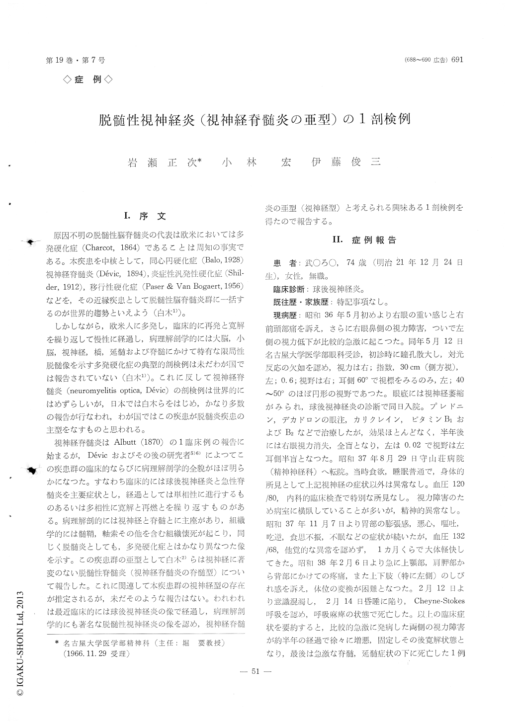
原因不明の脱髄性脳脊髄炎の代表は欧米においては多発硬化症(Charcot, 1864)であることは周知の事実である。本疾患を中核として,同心円硬化症(Balo, 1928)視神経脊髄炎(Devic, 1894),炎症性汎発性硬化症(Shil—der, 1912),移行性硬化症(Paser & Van Bogaert, 1956)などを,その近縁疾患として脱髄性脳脊髄炎群に一括するのが階界的趨勢といえよう(白木1))。
しかしながら,欧米人に多発し,臨床的に再発と寛解を繰り返して慢性に経過し,病理解剖学的には大脳,小脳,視神経,橋,延髄および脊髄にかけて特有な限局性脱髄像を示す多発硬化症の典型的剖検例は未だわが国では報告されていない(白木1))。これに反して視神経脊髄炎(neuromyelitis optica, Devic)の剖検例は世界的にはめずらしいが,日本では白木らをはじめ,かなり多数の報告が行なわれ,わが国ではこの疾患が脱髄炎疾患の主型をなすものと思われる。
A 74 year-old female was admitted with acute visual disturbance. She was diagnosed retrobulbar neuritis on examination of impaired visual acuity and optic atrophy on ocular fundi. After half a year, her visual acuity became to be 0 at right and 0.02 at left. Twenty months later, she complained of spontaneous pain on scapula and upper portion of back, intense "shibire" feeling, particularly on left upper and lower limbs. Thereafter, she died soon in coma and Cheyne-Stokes' respiration.
Marked gross and histopathological changes were fo-und mainly in the optic nerve. Severe demyelination, necrosis, glio-mesenchymal scarring and many cystic ca-vitation were noticed throughout entire area from the optic nerve to the ocular fundi. In these lesions, ven-ous vessels walls were fibrohyalinously thickened and perivascular lymphocytic infiltration occured in slight to moderate degree. Further, neurons of corpus ge-niculatum laterale were secondarily degenerated and in calcarine cortex, some eosinophilic bodies of unknown origin were scattered adjacent to capillaries.
Some localized lesions similar to those of the optic nerve as mentioned above were found in the white matter adjacent to the lateral ventricle. Slight in-flammatory infiltration developed in the lept-meninges.
In the center of upper cervical cord to the caudal most medulla oblongata, an acute softenning focus de-veloped.
Histopathological diagnosis of this case is demye-linating optic neuritis. The above histopathological findings seems to correspond to those of neuromyelitis optica, Devic. Because of minor change of spinal cord, this case is an atypical neuromyelitis optica, but seems to fall into the subtype (optic type).
The possible relation between the clinical retrobulbar neuritis and neuromylitis optica in Japan was discussed.

Copyright © 1967, Igaku-Shoin Ltd. All rights reserved.


