Japanese
English
- 有料閲覧
- Abstract 文献概要
- 1ページ目 Look Inside
I.緒言
今日におけるSturge-Weber病の通念は,Bergstrand,Olivecrona u.Tonnis (1936)が,その著"Über Ge—fässmissbildungen und Gefässgeschwülste des Gehirns"のなかで,既存の症例を,Sturge-Weber病の病名のもとに1つの単位疾患として包括したことに準拠している16)。
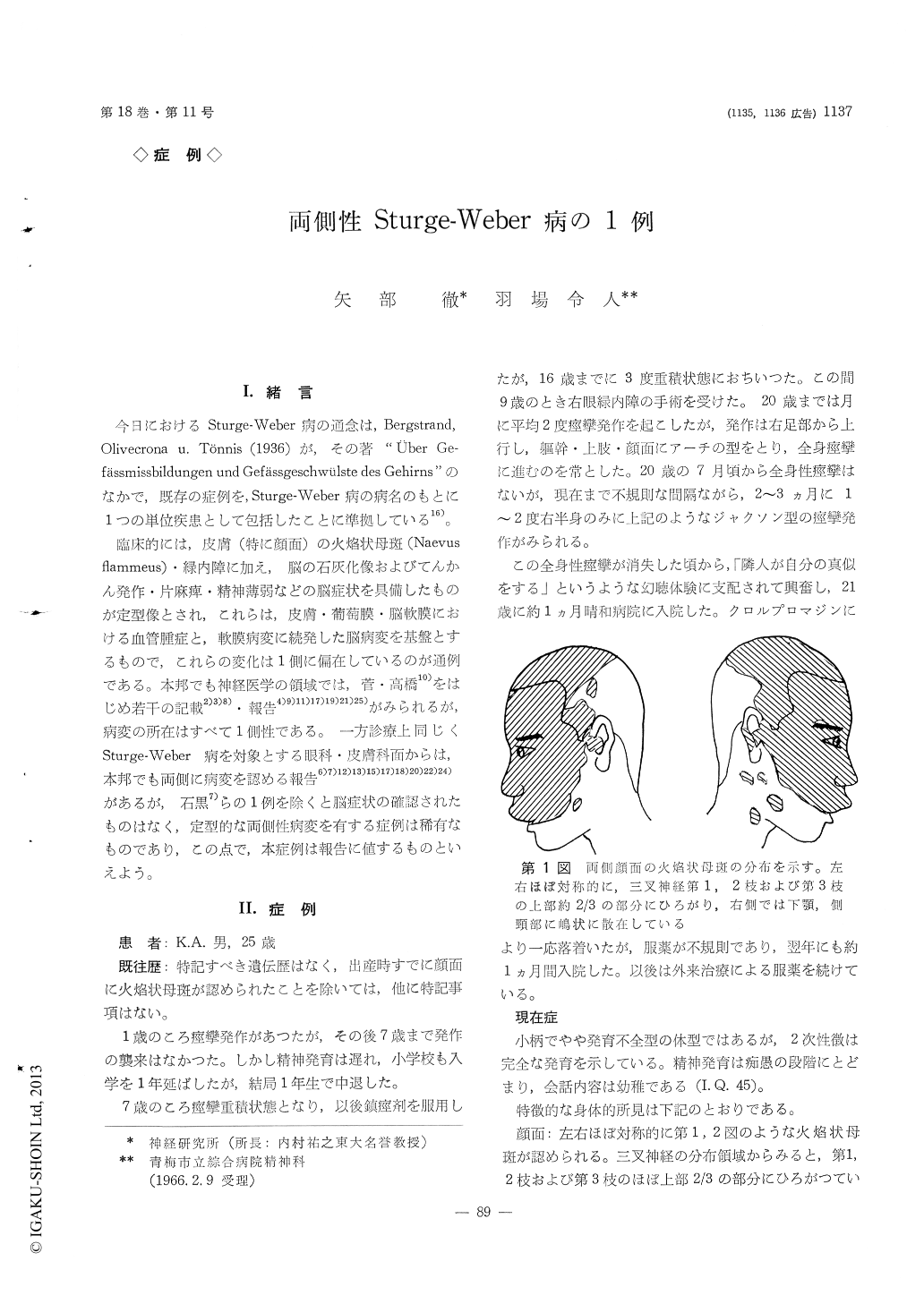
臨床的には,皮膚(特に顔面)の火焔状母斑(Naevus flammeus)・緑内障に加え,脳の石灰化像およびてんかん発作・片麻痺・精神薄弱などの脳症状を具備したものが定型像とされ,これらには,皮膚・葡萄膜・脳軟膜における血管腫症と,軟膜病変に続発した脳病変を基盤とするもので,これらの変化は1側に偏在しているのが通例である。本邦でも神経医学の領域では,菅・高橋10)をはじめ若干の記載2)3)8)・報告4)9)11)17)19)21)25)がみられるが,病変の所在はすべて1側性である。一方診療上同じくSturge-Weber病を対象とする眼科・皮膚科面からは,本邦でも両側に病変を認める報告6)7)12)13)15)17)18)20)22)24)があるが,石黒7)らの1例を除くと脳症状の確認されたものはなく,定型的な両側性病変を有する症例は稀有なものであり,この点で,本症例は報告に値するものといえよう。
A man, born in 1941.
There was nothing of note in the family history. Born at term, he was normal except for bilateral facial naevi flammei on the distribution area of 1 st, 2 nd and the upper two-thirds of 3 rd branch of the trigeminal nerve, almost symmetrically except small spots of naevi scattered in the mandible and lateral neck on the right side.
The mucosa of soft palate was hyperemic diffusely.
The conjunctiva in the both eyes were hyperemic, with an excess of fine vessels. The patient had an operation for right glaucoma at the age of nine, and lost the sight of right eye due to cataracts complicata. The left ocular tension showed 14. 5 mmHg, although optic disk was pale and excavated on its nasal side, demonstrated atrophy.
The first attack of convulsions was at the age of 12 months, and after an interval of six years convul-sions had appeared in Jacksonian type started from right foot.
Electroencphalographically the low voltage pattern was noticed, and a waves were observed only in right occipital region.
Radiological examination revealed string-shaped cla-cification located in the parieto-occipital portion on the both sides of the skull.
His mental development was retarded, and he could not help to leave school at the age of nine. The re-sults of the intelligence test revealed I. Q. 45.
From these findings, the bilateral Sturge-Weber Syndrome was confirmed.
Copyright © 1966, Igaku-Shoin Ltd. All rights reserved.