Japanese
English
- 有料閲覧
- Abstract 文献概要
- 1ページ目 Look Inside
I.緒言
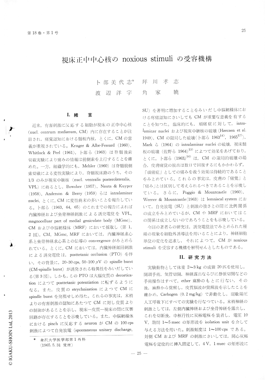
近来,有害刺激に反応する細胞が視床の正中中心核(nucl. centrum rnedianum, CM)内に存在することが注目され,痛覚認知における髄板内核,とくに,CMの意義が重視されている。Kruger&Albe-Fessard (1960),Whitlock&Perl (1961),卜部ら(1963)は脊髄後索切截実験により痛みの情報は前側索を上行することを確めた。一方,組織学的にも,Mehler (1960)は脊髄前側索切敵による変性実験により,脊髄視床路のうち,その1/3のみが視床中継核(nucl. ventralis posterolateralis, VPL)に終るとし,Bowsher (1957), Nauta&Kuyper(1958),Anderson&Berry (1959)らはintralaminar nuclei,とくに,CMに変性終末の多いことを報告している。卜部ら(1963,64,65)のこれまでの報告によれば内臓神経および坐骨神経刺激による誘発電位をVPL,magnocelluar part of medial geniculate body (MGmc),CMおよび中脳網様体(MRF)において採取し(第1,2図),CM, MGmc, MRFにおいては,内臓神経求心系と坐骨神経求心系との伝導のconvergenceがみとめられている。とくに,CMにおいては,内臓神経頻回刺激による誘発電位は,posttetanic occlusion (PTO)を伴い,その背景に,20-30 cps,50-100μVのspindle burst (CM-spindle burst)が誘発される特異性をみいだしている(第3図)。しかも,このPTOは大脳皮質のdecortica—tionによつてposttetanic potentiationに転ずるようになる。また,皮質のstrychnizationによつてCMにspindle burstを発現せしめ得た。これらの事実は,末梢よりの有害刺激の認知にあたつてCMに対し皮質よりの制御があることを示し,視床—皮質—視床の間に反響回路が存在することを示唆している。また,中脳網様体におけるpinchに反応するneuronがCMの100 cps刺激によつて自発放電(spontaneous unitary discharge, SU)を著明に増加することをみいだし中脳網様体における疼痛認知にさいしてもCMが重要な意義を有することを知つた。臨床的にも,頑痛症に対して,intra—lamlnar nucleiおよび視床中継核の破壊(Haecaen et al. 1949),CMの限局した破壊(卜部ら196314),196517)),Markら(1964)のintralaminar nucleiの破壊,視床髄板の破壊(佐野ら1964)13)によつて効果をあげており,とくに,卜部ら(1963.)14)は,CMの限局的破壊の場合,皮膚痛覚の脱出は数日で回復するにもかかわらず,「頑痛症」としての痛みを救う効果は持続的であろことをみとめている。これらの事実は,皮膚の「痛覚」と「痛み」とは区別して考えられるべきであることを示唆している。さらに,Poggio&Mountcastle (1960),Werner&Mountcastle (1963)はlemniscal systemにおいて,自発放電(SU)と刺激の強さとの間に比例関係の成立をみとめているが,CMやMRFにおいてはこの関係は成立しないのであろうことをも示唆している。
今回の著者らの研究は,誘発電位法でみとめられた種種の現象を細胞外誘導法を用いることにより,神経細胞単位の変化を追求し,それによつて,CMがnoxious stimuliを受容する機構を解明せんとしたものである。
Exploring microelectrodically the convergent neurons in nucleus centrum medianum (CM) of the thalamus of unanesthetized cats, which responded to sciatic and splanchnic stimulations as well as noxious stimuli, the uthors investigated the natures of these CM neurons induring sensory perception. The results are summariz-ed as follows :
1) The frequency of spontaneous unitary discharges (SU) from CM was 5 to 27 per second and showed relatively regular pattern of discharge. These spontane ous unitary discharges (SU) were markedly reduced and became grouped pattern, under light anesthesia. Following cortical strychnization or stimulation of contralateral CM and mid-brain reticular formation (MRF), spontaneous unitary discharges (SU) exhibited marked facilitation.
2) Follwing splanchnic and sciatic nerve stimula-tions unitary discharges (SU) were recorded with the average latencies of 18.7 and 18.0 msec, respectively. Following the tetanic stimulation (10 cps) to both nerves, driven unitary discharges (DU) disappeared and spontaneous unitary discharges (SU) were facilitated.
3) The pattern of facilitation of spontaneous dis-charges (SU) following the tetanic stimulation of the splanchnic nerve was formulated as four groupes : type 1 showed the neurons in close relation with spindle burst (16%), type 2 showed the neurons with facilitation during and after the tetanic stimula-tion (45%), type 3 showed the neurons with facili-tation during the tetanic stimulation and inhibition in a phasic modus after the tetanic stimulation (25%), and type 4 showed the neurons with an inconstant facilitation (16%).
4) Eighty two neurons in CM which were endowed with a full condititon described above, had two or more receptive field on both sides to the noxious stimuli.
5) Reactability of CM-neurons to the noxious stimuli was demonstrated by an increase of frequency of discharge and a prolongation of duration of disc-harge.
6) Reactibility of CM neuron to the noxious stimuli was augmented by the stimulation of MRF or contralateral CM.

Copyright © 1966, Igaku-Shoin Ltd. All rights reserved.


