Japanese
English
- 有料閲覧
- Abstract 文献概要
- 1ページ目 Look Inside
分類上の位置及び組織發生學的概念
所謂『上衣下細胞腫』が新しい範疇に屬する神經膠腫としてその分類のなかに導入されたのは1945年SCHE-INKR以來のことであり,その前後にあつては今日に至る迄全く記載されることがなかつた。腦腫瘍中,極端に稀な部類とは言い難い(SCHEINKER),而も非常に幅狹い分化の像を示す此の型の腫瘍が,今日までに斯うした名稱を冠して記載されてゐないのは,恐らく本型の部分像が全くastrocytomaに類似をもとめ得ることからその一異型としてか,あるいはenigmatic typeとして特別な注意を惹かされずに終つた結果であろうかと惟われる。1944年GLOBUS & KUHLENBECKは腦室周圍細胞床の特異な構造に着目して,胎生期より連續的に分化を示す上衣下グリアが,一定のremnis cent cell clus-terのかたちで上衣性腫瘍の展開に重要な場を占めるという事實を暗示し,續いて翌年その概念を裏附けるかの如くSCHEINKERは七例の定型的上衣下細胞腫を一括して,その特異の腫瘍學上の地位を體系づけるに至つたのである。私は之と全くその軌を一にした貴重な1例を教室剖檢例から檢索する機會を得たので茲に報告し,更に既定の記載を敷衍したいと思う。
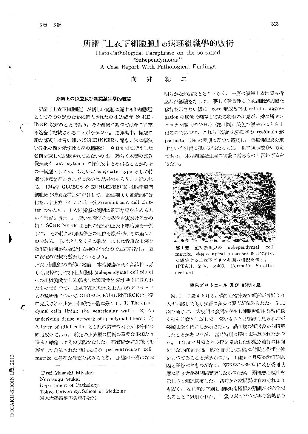
上衣下細胞腫の名稱は勿論,本型腫瘍が全く異型性に乏しく,顕著な上衣下性細胞床(subependyrnal cell pla e)への組織模倣をとる卓越した類同性を示すゆえに採られたものであつて,上衣下細胞母地と上衣型のグリオーマとの關聯性について,GLOBUS, KUBLENBECKは正常に完成された上衣下組織を三層に分つて,1) The epen—dymal cells lining the ventricular wall; 2) Anunderlying dense network of ependymal fibers; 3)A layer of glial cells.とし此の第三の因子が未分化の細胞成分であり,特定の上衣型の腫瘍の重要な根源たり得ると結論してその濫觴をなした。事實慥かに生後日を經ずして剖檢された新生兒腦のperiventricular cell matrixに仔細な觀察を試みるとき,上述の三層はなお明らかな形態をとることなく,一層の腦室上衣は?々折込んだ皺襞をなして,夥しく延長性の上衣細胞が明瞭な移行を示さない儘に,core形成乃至はcellular aggre—gationの状態で残存してゐる特有の所見が,殊に燐タングステン酸(PTAH.)(第1圖)染色で鮮やかにとらえ得るのであつて,これら原始的未熟細胞のresidualsがpostnatal lifeの長期に亙つて遺殘し,腫瘍性變異を來すとい事實に狙いを得たことは,兎に角示唆多い考えであり,本型組織發生論の肯綮に當るものと言わざるを得ない。
There is general agreement that subependy-moma is a distinct tumor entity since the des-criptions of Globus and Kuhlenbeck (1944) and Scheinker (1945). This terminology is still un-common in Japan and, in fact, the tumor is rarely encountered, because the attention has not been directed to the possibility of tumor derivation from the subependymal neuroglial elements. In the author's serial necropsy cases of gliomas, one subependymoma was found.
CASE REPORT: This patient was first seen at the age of five years with a high fever and in coma. His first similar seizure occurred at the age of one year and he had had recurrence at about monthly intervals. Associated with this were periodic changes, listlessness and fatigability, and he gradually developed speech difficulties. At the age of three years his parents stated that he had developed difficulty in walking, which by the age of four years had become quite marked and he had developed a bilateral pes equinovarus. On his admission to the hospital at the age of five years he was in coma and had a spastic paralysis of the left half of the body and had grand mal epileptic seizures frequently. He had bilateral papille-dema. The cerebro-spinal fluid was clear with an initial pressure of 600 mm. of water. It con-tained 45/3 (Fuchs-Rosenthal) of cells and 150-mg/dl. of sugar. Diagnostic pneumoencephalo-grams were made, following which he went into shock and died.
Post Mortem Findings: There was a fist-sized semitransparent grayish-brown, firm tumor in-volving the pons and obliterating the lumen of the fourth ventricle and the aqueduct of Sylvius. Microscopic appearance of the tumor was identical to that described by Scheinker in 1945. The stroma was ectodermal origin, no mesen-chymal reaction was seen. This tumor is deriv-ed from the subedendymal neroglial elements and thus assumes the character of mature gli-al tissue. Scheinker described that the tumor constitute a well defined group of gliomas with the following salient characteristics: (a) The predominant cell is a mature fibrillary astrocy-te displaying a cell morphology and cell arrange-ment which duplicate the subependymal glia.(b) There is great preponderance of glial fibers over cellular elements. (c) The tumor is well demarcated from the surrounding tissue, reveal-ing thus an expansive type of growth rather than an infiltrative growth. Serial sections (3.0 by 5.0) by 103 by 103 microns of the tumor dis-closed this characteristic cellular make-up throughout. Thorough cytological investigationsof this type of tumor, particularly in its dis-tinctive staining properties and the argentaffin metallic impregnability, displayed a definite close kinship between subependymal glia and ependymal cells.
As a result of his study of this tumor, it is the author's belief that the opinion expressed by Willis and Cox that too much stress has been placed on the presence of blepharoplasten in ependymal cells is correct. The author has previously expressed this conviction. My studies suggest the hypothesis that miscellaneous type of gliomata, astrocytoma and epedymoblastoma may originate from a common stem (primordi-al) cell.

Copyright © 1953, Igaku-Shoin Ltd. All rights reserved.


