Japanese
English
- 有料閲覧
- Abstract 文献概要
- 1ページ目 Look Inside
ホースレイ,クラークが動物腦深部に障害を與える方法を考案して以來長い間經つたが,人間では頭蓋に個人差が多いので今まで行われなかつた。著者は之を人間に行う装置を考案した。即ち石灰化した松果體或いは腦室像を目標にして侵襲部位の位置的關係を正確にきめ,電極を小さな穿顱孔から刺入して,必要な部分に限局した障害を加えるのである。
1) 精神外科的應用。精神病に對してタラモトミー,ヒボタラモトミーを行う。
In various diseases of the central nervous system, is desirable to carry out therapeutic procedures in the depth of the brain. The classical methods of brain surgery necessitate the exposure of such deep lying areas. Usually this is associated with unavoidable damage to parts of the brain located above the region which the surgeon attempts to reach. However, over 40 years ago a method had been introduced by Horsley and Clark1) for experimental work on animals which permits one to place lesions in deep-seated areas of the brain with minimum injury to overlying structures. Curiously enough, as far as we know, this method has not been applied to humans prior to our first publi—cation2) (Sience 106: 349, Oct. 10, 1937). The main reason for the previous limitation of this method to experimental animals is the great variability in size and shape of the skull in man. This makes it impossible to place a lesion in a certain subcortical area with any degree of accuracy if one uses a landmark of the skull as a reference point, such as, for instance, the external auditory meatus. This is possible in such animals only in which size and shape of the head are rather uniform such as, for instance, in cats and monkeys. We have tried to overcome these difficulties regarding the human skull by using a point within the brain, usually the pineal body, as a reference point. Its position in relation to the coordinates of the apparatus is determined by simple x-ray photo—graphy of the skull with the apparatus insitu if the pineal gland is calcified. Otherwise visualization of the ventricles by pneumoence—phalography is necessary.
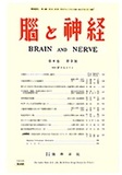
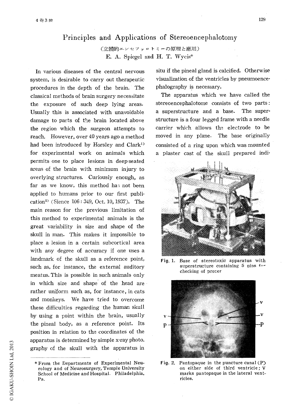
The apparatus which we have called the stereoencephalotome consists of two parts: a superstructure and a base. The super—structure is a four legged frame with a needle carrier which allows the electrode to be moved in any plane. The base originally consisted of a ring upon which was mounted a plaster cast of the skull prepared indi—vidually for each patient. A newer modifi—cation (Fig. 1, 2) climinates the plaster cast and anchors the base of the ste eoence—phalotome to the skull by means of four rubber plugs or by four stainless steel pins which are inserted into small drill holes of the skull. The base of the apparatus is aligned so that its frame is parallel to a plane deter—mined by the inferior border of the orbits and the external auditory canals. the so-called horizontal plane. As already mentioned, the coordinates of the apparatus are referred to a point within the brain, namely the pineal gland. The patient is x-rayed with the apparatus in position and the relationship of the pineal gland to the electrode is establi—shed.
Copyright © 1952, Igaku-Shoin Ltd. All rights reserved.