Japanese
English
- 有料閲覧
- Abstract 文献概要
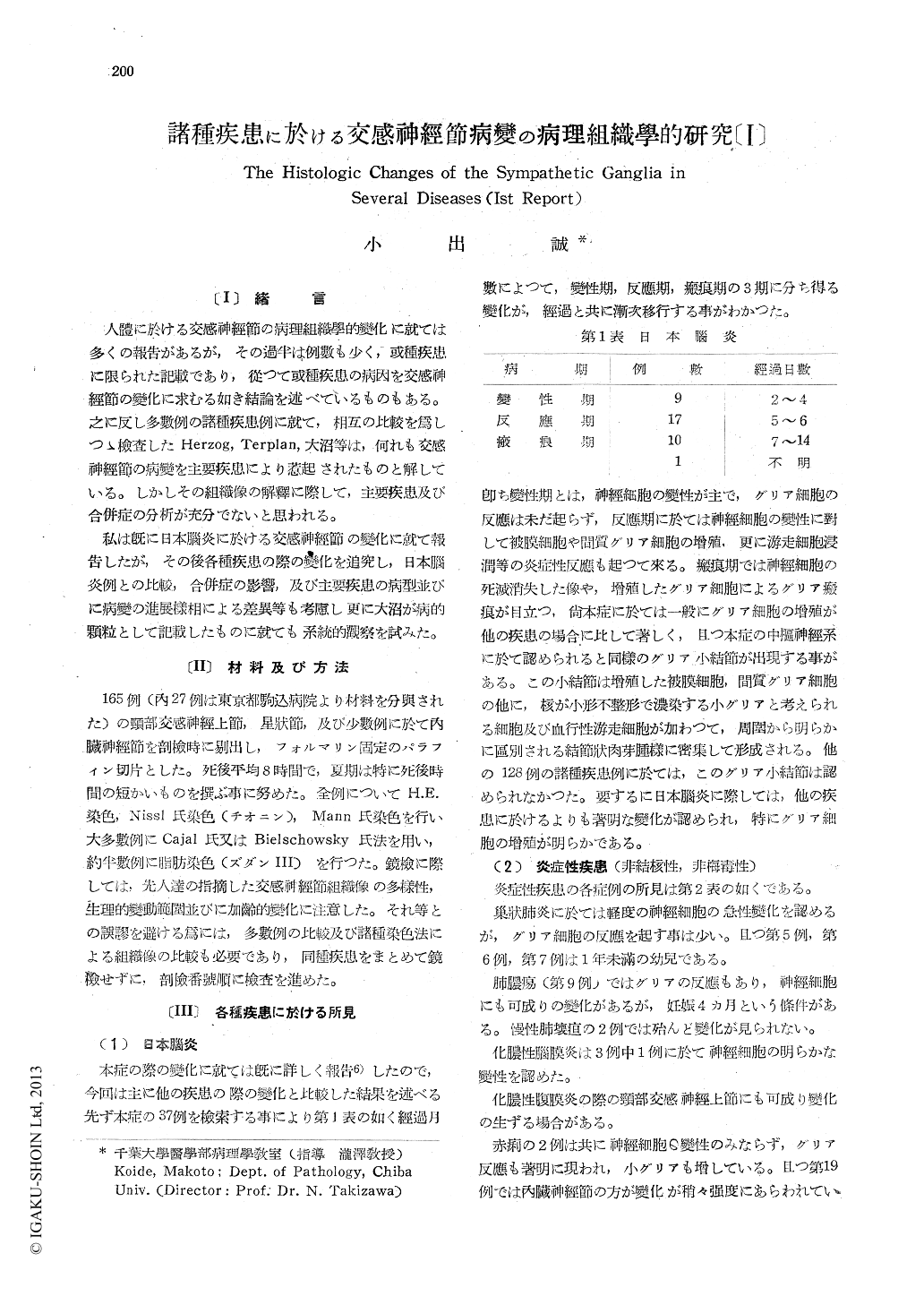
- 1ページ目 Look Inside
〔1〕緒言
人體に於ける交感神經節の病理組織學的變化に就ては多くの報告があるが,その過半は例數も少く,或種疾患に限られた記載であり,從つて或種疾患の病因を交感神經節の變化に求むる如き結論を述べているものもある。之に反し多數例の諸種疾患例に就て,相互の比較を爲しつゝ檢査したHerzog, Terplan,大沼等は,何れも交感神經節の病變を主要疾患により惹起されたものと解している。しかしその組織像の解釋に際して,主要疾患及び合併症の分析が充分でないと思われる。
私は既に日本腦炎に於ける交感神經節の變化に就て報告したが,その後各種疾患の際の變化を追究し,日本腦炎例との比較,合併症の影響,及び主要疾患の病型並びに病變の進展樣相による差異等も考慮し更に大沼が病的顆粒として記載したものに就ても系統的觀察を試みた。
By microscopic examination on the Ganglion cerv. sup. and Ggl. stell. (and Ggl. coel, in a few cases) in 165 cases, the following results were obtained.
1) Japanese B encephalitis.
I already reported the findings in this dise-ase. (Jap. Med. Jour. Vol. 3, No. 2)
The changes can be divided into 3 stages, namely the stage of degeneration, reaction, and cicatrix, according to the time from the onset of the illness. The proliferation of glia cells are seen more evidently than in other diseases, and the characteristic finding is the "neurog-lial nodulel."
2) Inflammatory dseases (non tuberculous,non syphilitic).
In bronchopneumonia, there is the degenera-tion of the nerve cells, while, in chronic gan-grene of the lung almost no changes are found.
In bacillary dysentery, there were noted not only the degeneration of the nerve cells but also the react.ons of the glia cells,
In some cases severe changes were found in the Ggl. coel. rather than in cervical ganglia. In septic diseases acute degeneration (subacute or chronic, in the case of endocarditis lenta) of the nerve cells were noted, complicated occaisionally by the proliferation of the glia cells, the edema of interstitium, cell-infiltra-tion, or bleeding.
3) Tuberculous disease.
In pulmonary phthisis with mainly produc-tive foci there was no change in those ganglia. In the cases with wide exudative foci, especi-ally in caseous pneumonia, there occured the changes in those gang lia. Tubercles were found in these ganglia in 3 of 33 tuberculous cases.
4) Syphilitic diseases.
Almost no changes in the nervous tissue of the sympathetic ganglia were found. But occ-asionolly existed the perivascular cell-infiltra- tion.
5) Broken metabolism and intoxication
Various degree of degeneration of the nerve cells were observed.
6) Diseases of blood.
Findings are the bleeding in the interstitium in purpura and evident infiltration of the myelogenic cells in myelogenic leucemia.
7) Malignant tumor.
When the case is not accompained by any complications, we can find hardly any changes. Therefore, I consider, the changes which exist in some cases with complications can be att-ributed to the influence of the complications namely pneumonia, suppurative perito-nitis, icterus, sepsis, etc.
Findings common to all cases. The nerve cells show changes according to various stages of degeneration. The cells melt-ing away, leave the amorphous or reticular vestiges of them. But at last they completely disappear and the lumen of the capsule become empty. The vacuoles are often noted, and they react negatively to fatstains (Sudan Ⅲ). Reacting to the degeneration of the nerve cells, the capsular cells swell or proliferate, and neuronophagia is occasionally noted.
The capsular cells surrounding the lumen aggregate centripetally and form the glial cicatrix, which corresponds to "Knotchen" des-cribed by Terplan and not to the "neuroglial nodule" in Japanese B encephalitis. On the wall of blood-vessels or perivascular connective tissue, we see the fibrous or hyalinous thickn-ing, as the age of patient advances, and round-cell-infiltration mostly accompanied by the changes of nerve cells. Bleeding could be seen in ten cases of all cases. The relationship bet-ween the location of main foci and the position of injured sympathetic ganglia is still obs-cure.

Copyright © 1951, Igaku-Shoin Ltd. All rights reserved.


