Japanese
English
- 有料閲覧
- Abstract 文献概要
- 1ページ目 Look Inside
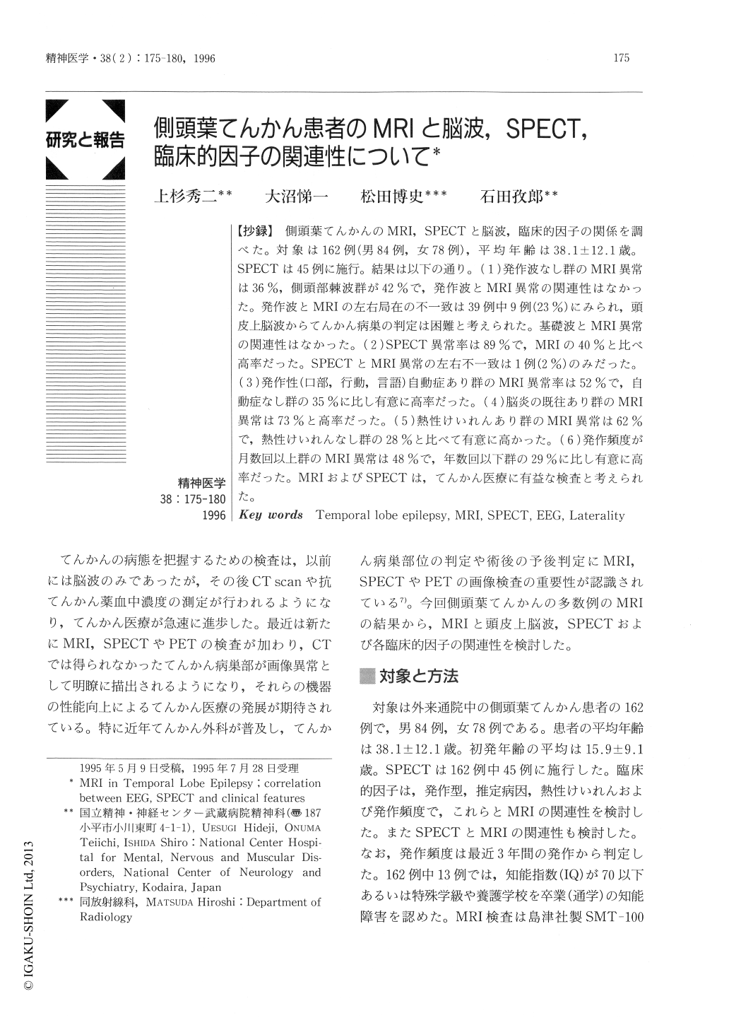
【抄録】側頭葉てんかんのMRI,SPECTと脳波,臨床的因子の関係を調べた。対象は162例(男84例,女78例),平均年齢は38.1±12.1歳。SPECTは45例に施行。結果は以下の通り。(1)発作波なし群のMRI異常は36%,側頭部棘波群が42%で,発作波とMRI異常の関連性はなかった。発作波とMRIの左右局在の不一致は39例中9例(23%)にみられ,頭皮上脳波からてんかん病巣の判定は困難と考えられた。基礎波とMRI異常の関連性はなかった。(2)SPECT異常率は89%で,MRIの40%と比べ高率だった。SPECTとMRI異常の左右不一致は1例(2%)のみだった。(3)発作性(口部,行動,言語)自動症あり群のMRI異常率は52%で,自動症なし群の35%に比し有意に高率だった。(4)脳炎の既往あり群のMRI異常は73%と高率だった。(5)熱性けいれんあり群のMRI異常は62%で,熱性けいれんなし群の28%と比べて有意に高かった。(6)発作頻度が月数回以上群のMRI異常は48%で,年数回以下群の29%に比し有意に高率だった。MRIおよびSPECTは,てんかん医療に有益な検査と考えられた。
The relationship between MRI, SPECT, EEG and clinical features in temporal lobe epilepsy was investigated. Subjects were 162 patients (84 males, 78 females) whose average age was 38.1±12.1 years. SPECT was carried out in 45 patients.【Results】 (1) Abnormal MR images were obtained in 36% of the group without epileptic discharge, and in 42% of the group with temporal spikes. There was no correlation between epileptic discharge in EEG and MRI abnormality. The lateralities of epileptic discharge and MRI were in disagreement in 9 of 39 patients (23%), indicating that determining the epileptic focus from scalp EEG was difficult. There was no correlation between the basic activity in EEG and abnormality in MRI. (2) The rate of abnormal SPECT (89%) was higher than that of abnormal MRI (40%). (3) The rate of the group with ictal automatism (52%) was higher than that of the group without ictal automatism (35%). (4) The rate of abnormal MR images was high in the group with encephalitis (73%). (5) The rate was higher in the group with febrile convulsion (62%) than in the group without it (28%). The rate of the abnormal MR images was higher in the group with a seizure frequency of at least several mal/ month (48%) than in the group with a seizure frequency of less than several mal/year (29%).

Copyright © 1996, Igaku-Shoin Ltd. All rights reserved.


