Japanese
English
- 有料閲覧
- Abstract 文献概要
- 1ページ目 Look Inside
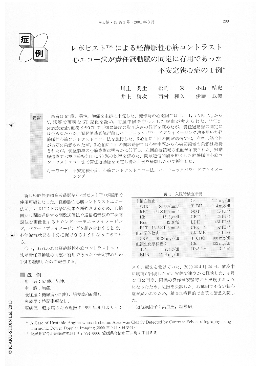
要旨 患者は67歳,男性.胸痛を主訴に来院した.発作時の心電図ではI,II,aVF,V2からV6誘導で著明なST変化を認め,前壁中隔を中心とした虚血が考えられた.99mTc-tetrofosmin血流SPECTで下壁に軽度の取り込みの低下を認めたが,責任冠動脈の同定には至らなかった.冠動脈造影施行前にハーモニックパワードプライメージング法を用いた経静脈性心筋コントラストエコー法を施行した.6心拍に1回の問激送信では,左室心筋全体が良好に染影されたが,3心拍に1回の間歇送信では心室中隔から心尖部領域の染影は維持されたが,側壁領域の心筋染影は明らかに低下し,左回旋枝領域の虚血が示唆された.冠動脈造影では左回旋枝#11に90%の狭窄を認めた.間歌送信間隔を短くした経静脈性心筋コントラストエコー法で責任冠動脈を同定し得た1例を経験したので報告した.
A 67-year-old man was admitted to our hospital dueto unstable angina. Electrocardiography at rest revealedST segment depression in II, aVF, and V4 to V6 andfurther depression was observed during chest pain. 99mTc-tetrofosmin SPECT demonstrated perfusion defect atthe inferior area. We performed harmonic power Doppler imaging with contrast echocardiography using adigital ultrasound system (SYSTEM FIVE, Vingmed, Norway). Initially, harmonic power Doppler imagingwas obtained by intermittent (one per 6 cardiac cycles)imaging. Perfusion defect was detected neither at theapical four chamber view nor at the two chamber view.To decrease the supply of microbubbles, we changed theintermittent rate from one per six to one per threecardiac cycles. It was then that the perfusion defect inthe lateral area was clearly detected at the apical fourchamber view. Coronary arteriography revealed 90%stenosis at the mid left circumflex artery. After percutaneous transluminal coronary angioplasty at the midcircumflex artery, the perfusion defect in the lateralarea was disappeared and normalized. Harmonic powerDoppler imaging by contrast echocardiography wasuseful for detecting the ischemic area in a patient withunstable angina.

Copyright © 2001, Igaku-Shoin Ltd. All rights reserved.


