Japanese
English
- 有料閲覧
- Abstract 文献概要
- 1ページ目 Look Inside
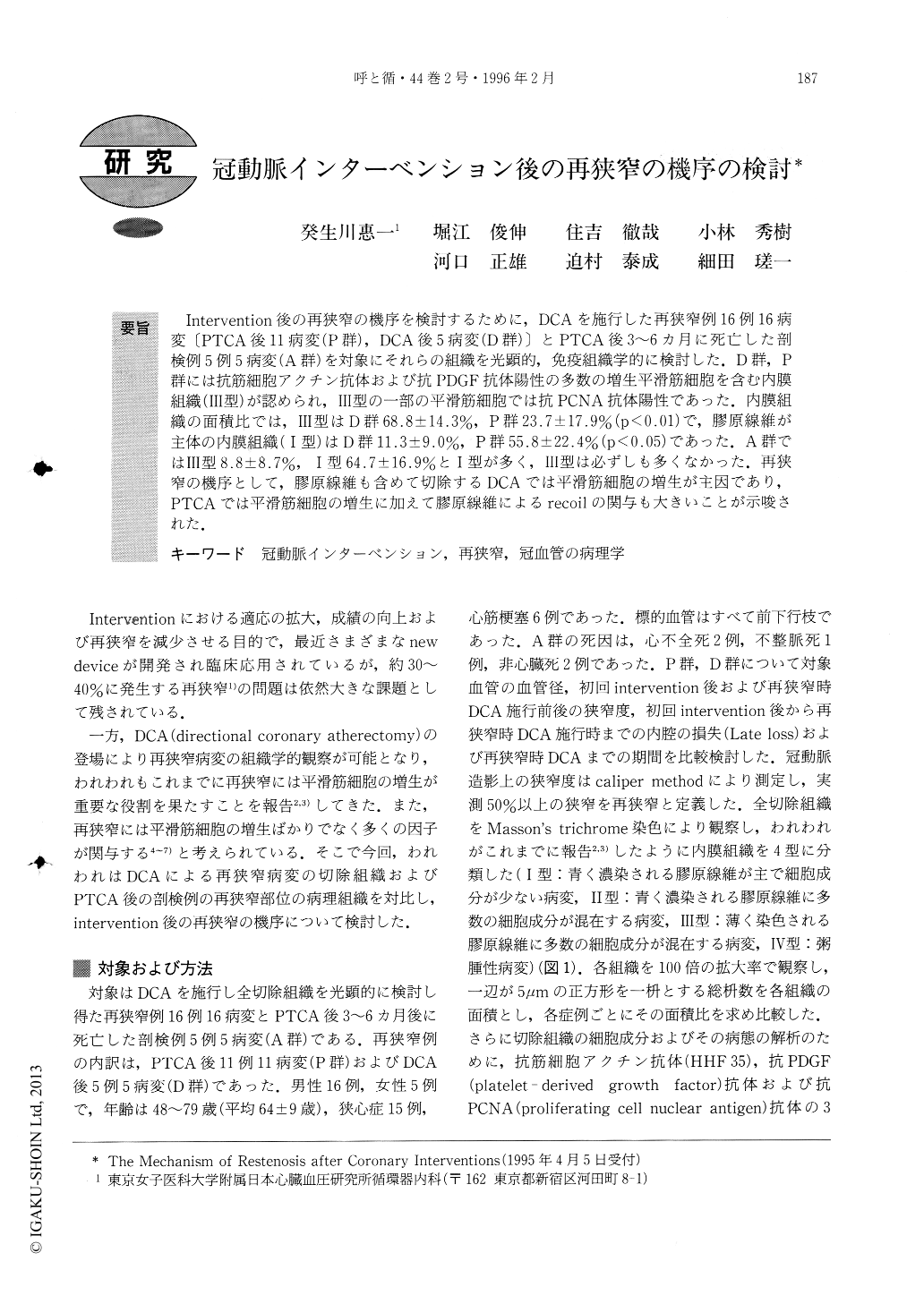
Intervention後の再狭窄の機序を検討するために,DCAを施行した再狭窄例16例16病変〔PTCA後11病変(P群),DCA後5病変(D群)〕とPTCA後3〜6カ月に死亡した剖検例5例5病変(A群)を対象にそれらの組織を光顕的,免疫組織学的に検討した.D群,P群には抗筋細胞アクチン抗体および抗PDGF抗体陽性の多数の増生平滑筋細胞を含む内膜組織(III型)が認められ,III型の一部の平滑筋細胞では抗PCNA抗体陽性であった。内膜組織の面積比では,III型はD群68.8±14.3%,P群23.7±17.9%(p<0.01)で,膠原線維が主体の内膜組織(I型)はD群11.3±9.0%,P群55.8±22.4%(p<0.05)であった.A群ではIII型8.8±8.7%,I型64.7±16.9%とI型が多く,III型は必ずしも多くなかった.再狭窄の機序として,膠原線維も含めて切除するDCAでは平滑筋細胞の増生が主因であり,PTCAでは平滑筋細胞の増生に加えて膠原線維によるrecoi1の関与も大きいことが示唆された.
We investigated the mechanism of restenosis after coronary interventions. Sixteen patients who had 16 restenotic lesions, of which 11 lesions (group P) were after percutaneous transluminal coronary angioplasty (PTCA) and 5 lesions (group D) were after directional coronary atherectomy (DCA), underwent DCA. All resected tissues from group P, group D and necropsied coronary arteries from 5 patients who died 3-6 months after PTCA (group A) were examined histopath-ologically and immunohistochemically.
No significant difference between group P and group D were found in baseline quantitative angiographic variables (mean vessel size 3.46±0.73mm versus 3.49±0.72mm and preprocedural percent diameter stenosis 81±12% versus 86±14%). Significant difference was found in the percent diameter stenosis after prior inter-vention (group P versus group D=27±7% versus 8±4%; p<0.01), whereas the preprocedural percent diam-eter stenosis of group P was similar to that of group D (84±8% versus 79±9%) at secondary intervention (DCA). Late lumen loss in group D was larger than that in group P. Because the percent diameter stenosis after DCA of group P and group D was similar (10± 10% versus 6±10%), most tissues of the restenotic lesions of the two groups were similarly resected by DCA.
Resected intimal tissues stained with Masson's tri-chrome were classified into four groups according to the characteristic fibrous tissue and the amount of prolifer-ative cells: Type I= old dense fibrous tissue, Type II= relatively old fibrous tissue containing many prolifer-ative cells, Type III= new loose fibrous tissue contain-ing many proliferative cells, and Type IV=ather-omatous plaques. The proliferating cells of the Type III tissues stained positive with HHF35 (anti-muscle actin monoclonal antibody) and anti-PDGF (platelet-derived growth factor) monoclonal antibody, and some of these cells stained positive with anti-PCNA (proliferating cell nuclear antigen) monoclonal antibody. Therefore, the Type III tissues contained “active” smooth muscle cells and the Type III tissues were “active” intimal tissues. Type III tissues occupied 68.8±14.3% of the area of all resected intimal tissues in group D and 23.7±17.9% in group P (p<0.01). On the contrary, Type I tissues occupied 11.3±9.0% in group D and 55.8±22.4% in group P (p<0.05). In group A, Type III tissues were shown in 8.8±8.7% and Type I tissues in 64.7±16.9%.
We found that recurrent stenosis after DCA which can excise coronary artery lesions including fibrous tissues was due mainly to proliferation of smooth muscle cells, and restenosis after PTCA was due not only to proliferation of smooth muscle cells but also to elastic recoil of fibrous tissue.

Copyright © 1996, Igaku-Shoin Ltd. All rights reserved.


