Japanese
English
- 有料閲覧
- Abstract 文献概要
- 1ページ目 Look Inside
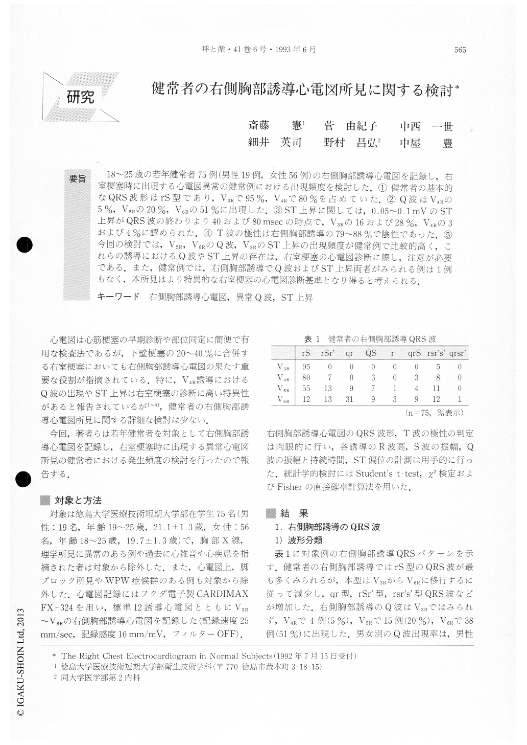
18〜25歳の若年健常者75例(男性19例,女性56例)の右側胸部誘導心電図を記録し,右室梗塞時に出現する心電図異常の健常例における出現頻度を検討した.①健常者の基本的なQRS波形はrS型であり,V3Rで95%,V4Rで80%を占めていた.②Q波はV4Rの5%,V5Rの20%,V6Rの51%に出現した.③ST上昇に関しては,0.05〜0.1mVのST上昇がQRS波の終わりより40および80 msecの時点で,V3Rの16および28%,V4Rの3および4%に認められた.④T波の極性は右側胸部誘導の79〜88%で陰性であった.⑤今回の検討では,V5R,V6RのQ波,V3RのST上昇の出現頻度が健常例で比較的高く,これらの誘導におけるQ波やST上昇の存在は,右室梗塞の心電図診断に際し,注意が必要である.また,健常例では,右側胸部誘導でQ波およびST上昇両者がみられる例は1例もなく,本所見はより特異的な右室梗塞の心電図診断基準となり得ると考えられる.
Right chest electrocardiograms (ECGs) of 75 healthy subjects (19 men and 56 women ; age 18 to 25 years) were recorded in order to study characteristic features of right precordial ST-T and QRS waves. The diagnostic criteria for right ventricular infarction (RVI) by ECG were also tested in healthy subjects to reascertain their value for diagnosing RVI. 1) The QRS configuration in right chest leads was usually the rS pattern in normal subjects (95% of V3R. and 80 % of V4R). 2) The Q wave was found in 5 % of V4R, 20% of V5R. and 51% of V6R.

Copyright © 1993, Igaku-Shoin Ltd. All rights reserved.


