Japanese
English
- 有料閲覧
- Abstract 文献概要
- 1ページ目 Look Inside
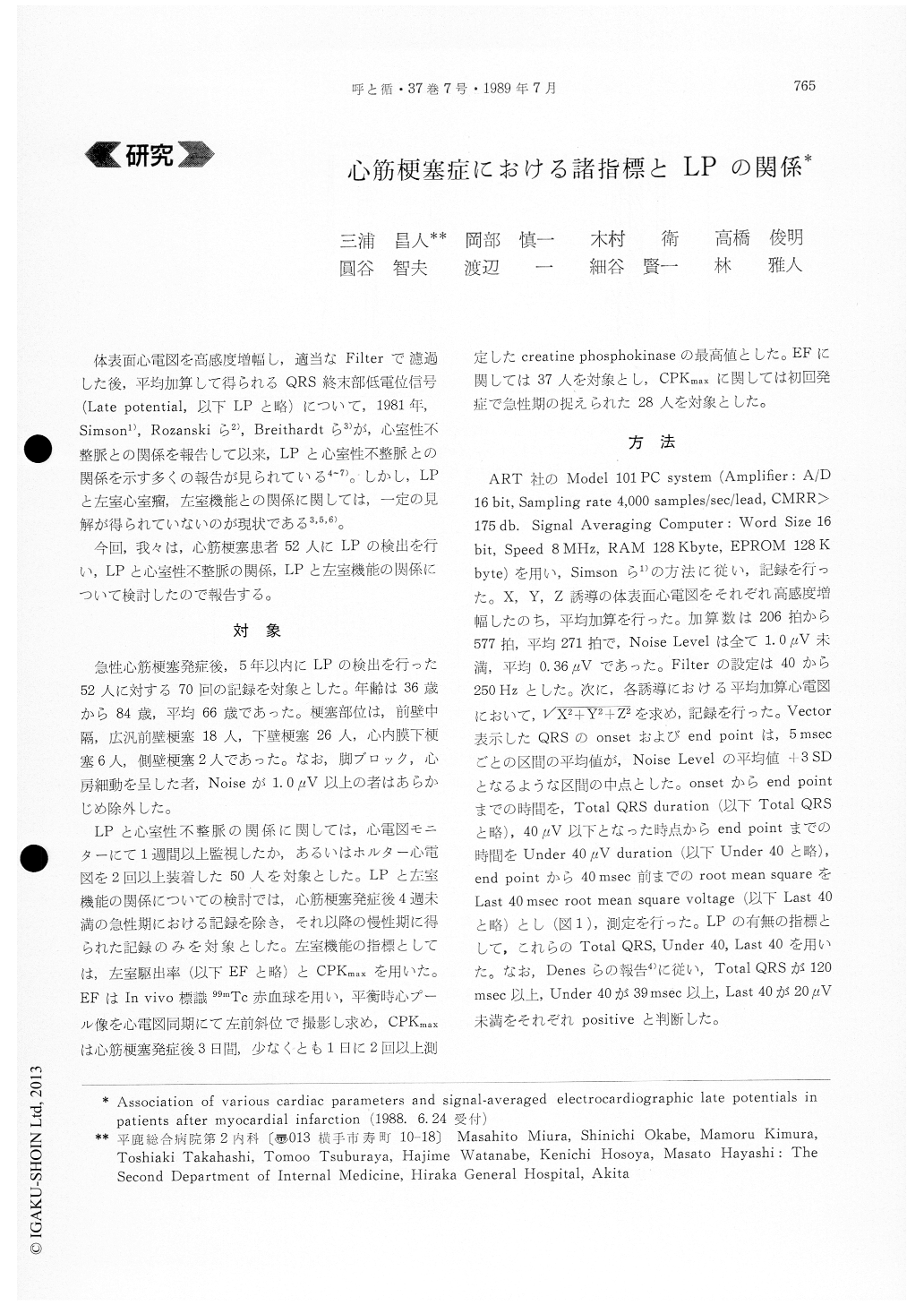
体表面心電図を高感度増幅し,適当なFilterで濾過した後,平均加算して得られるQRS終末部低電位信号(Late potential,以下LPと略)について,1981年,Simson1),Rozanskiら2),Breithardtら3)が,心室性不整脈との関係を報告して以来,LPと心室性不整脈との関係を示す多くの報告が見られている4〜7)。しかし,LPと左室心室瘤,左室機能との関係に関しては,一定の見解が得られていないのが現状である3,5,6)。
今回,我々は,心筋梗塞患者52人にLPの検出を行い,LPと心室性不整脈の関係,LPと左室機能の関係について検討したので報告する。
After the amplification and filteration of the sur-face ECG, late potential signal (LP) in QRS termina-tion was determined. Subjects were 52 patients who experienced acute myocardial infarction within 5 years and measurement was undertaken 70 times. 271 pulses (average) were measured using the Model 101 PC system (ART Co, Ltd) with 40-250 Hz filter. Relationship of LPs and ventricular tachyarrhyth-mia, LPs and left ventricular function were studied according to the ejection fraction (EF) of left ven-trium (=index of left ventricular function) and ma-ximum value of CPK (CPKmax) in acute phase. The patients with bundle branch block, atrial fibrillation and noise above 1.0μV were excluded.
Results : (1) Higher incidence of LP was shown in patients with triplet or more runs of ventricular tachycardias. (2) Higher incidence of LP was shown in patients with lower EF and higher CPKmax.
Above results showed that non-invasive and simple recording of LPs in patients with myocardial infarc-tion was useful in the discrimination of the patients with ventricular arrhythmia and presumption of left ventricular function.

Copyright © 1989, Igaku-Shoin Ltd. All rights reserved.


