Japanese
English
- 有料閲覧
- Abstract 文献概要
- 1ページ目 Look Inside
右側大動脈弓に発生した解離性大動脈瘤は非常に稀な疾患である。我々が文献的に検索した限りでは,世界で今までに4例報告されているのみである。本邦では,本症例が2例目であるの本症例は,右胸水貯留があったため初期は診断が困難であったが,CT心エコーにより解離性大動脈瘤(De Bakey III型)と診断することができた。本症例は,先天性心疾患の合併やチアノーゼもなく,右側大動脈弓の一般的なタイプと考えられた。本症例は,入院時は大動脈解離のため,プレショック状態であったがその後は内科的治療のみにて経過良好で無事退院となった。その臨床経過および若干の文献的考察を加え報告する。
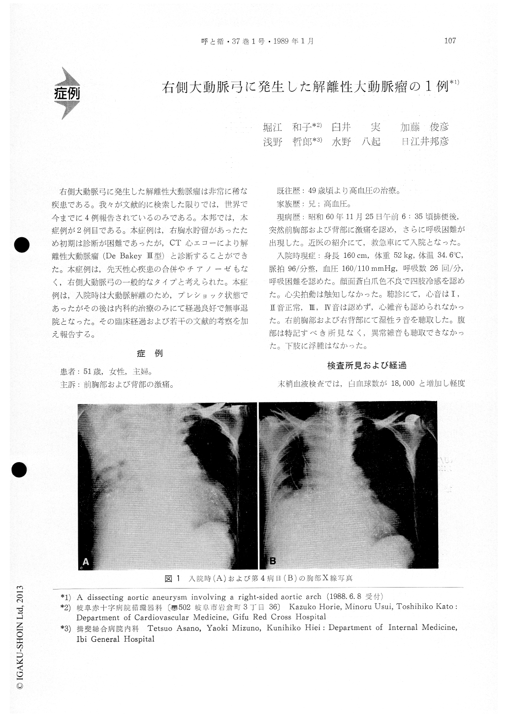
A 51-year-old woman suddenly developed severe pain in the chest and back, and also dyspnea. On admission, she was in a state of preshock. Plain X-ray indicated the lack of the left aortic arch and poor pneumatization in the whole right lung. The thoracic fluid was transparent with yellowish tinge and was contaminated with neither any bacterium nor tubercle bacillus. The response to the Rivalta's reaction was negative. The possibility of pleurisy was, therefore, denied. The ECG and blood bioch-emical data on the second day suggested the pos-sibility of myocardial ischemia.
Plain chest X-ray on the fourth day revealed an increased right pulmonary pneumatization and an enlarged mediastinal shadow toward the aortic arch. Upper pulmonary CT showed a mass on the right side. Enhanced CT disclosed a dissepiment in the center, which was high medially and somewhat lowlaterally. It was diagnosed as a false lumen due to the lateral displacement of the right aortic arch. Hepatic CT disclosed the tapering of the abdominal aorta from right to left in the prevertebral region. These findings indicated that the aorta descended from the right aortic arch along the right side of the spine and crossed the spine dextrosinistrally at the heptic level.In addition, dissociant aneurysm was observed in the right aortic arch. Echocardiography showed no evidence of dissociant aneurysm at the aortic base. Chest X-ray, CT and echocardiography showed the dissociation of the aorta from the aortic arch to the abdominal aorta. Thus the diagnosis of De Bakey type was established. Clinically, DIC and multiorgan disorders were manifested but after medical treatments, the clinical course was une-ventful.
The incidence of right-sided aortic arch is gener-ally estimated to be approximately 0.1%. There are two major anatomical types of right-sided aortic arch. One is a mirror image diametrical to the normal, having three vessels from the aortic origin. Cyanotic congenital heart diseases may occur in most patients with this type. The other is the most common type with four vessels from the aortic base. The present case seems to belong to the lat-ter type.
Dissociant aneurysm involving the right-sided aortic arch is extremely rare. To the best of our knowedge, this is the fifth reported in the whole world and the second in Japan.

Copyright © 1989, Igaku-Shoin Ltd. All rights reserved.


