Japanese
English
- 有料閲覧
PPV購入案内
1,320円 (1,200円+税10%) こちらは、470ページから476ページの文献です。購入には医書.jp本会員登録が必要です。
会員登録完了後に改めて上記「購入サイトへ」を選択してください。
または「購入サイトへ」選択後に「購入ログイン」、「新規会員登録」の順に進んでください。
会員登録について詳しくはをご確認ください。
- Abstract 文献概要
- 1ページ目 Look Inside
- 参考文献 Reference
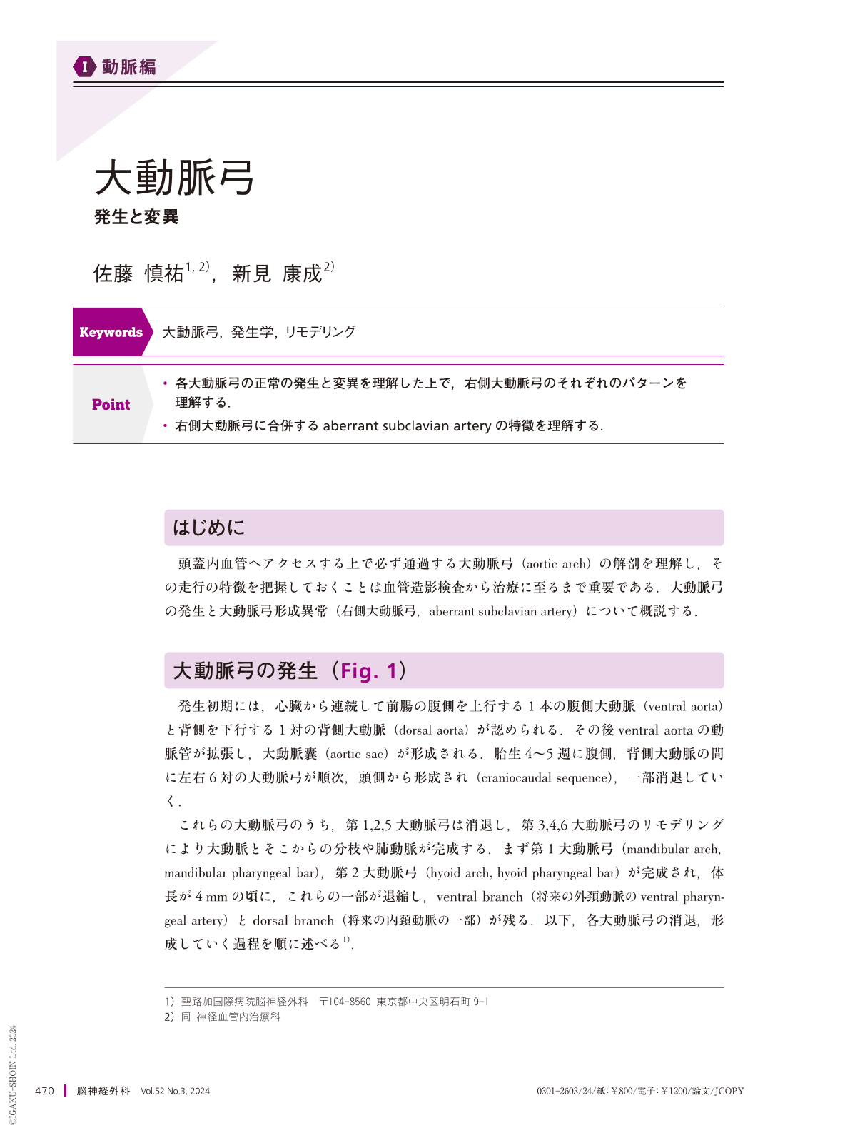
Point
・各大動脈弓の正常の発生と変異を理解した上で,右側大動脈弓のそれぞれのパターンを理解する.
・右側大動脈弓に合併するaberrant subclavian arteryの特徴を理解する.
A right aortic arch and aberrant subclavian artery result from an interruption in the remodeling of the pharyngeal arch arteries. We occasionally encounter this anatomical variation during angiography. Patients with disorders such as Down syndrome and congenital heart disease show a high incidence of an aberrant right subclavian artery, and this anomaly can cause symptomatic esophageal or tracheal compression. The root of the aberrant artery may show dilatation(referred to as a Kommerell diverticulum), dissection, intramural hematoma, or rupture necessitating cardiac intervention using a surgical or endovascular approach. Neurointerventionalists should have working knowledge of the anatomy to rapidly understand the anatomy and ensure a safe procedure. A left transradial approach should be considered if prior knowledge of the aberrant subclavian anatomy is available.
Copyright © 2024, Igaku-Shoin Ltd. All rights reserved.