Japanese
English
- 有料閲覧
- Abstract 文献概要
- 1ページ目 Look Inside
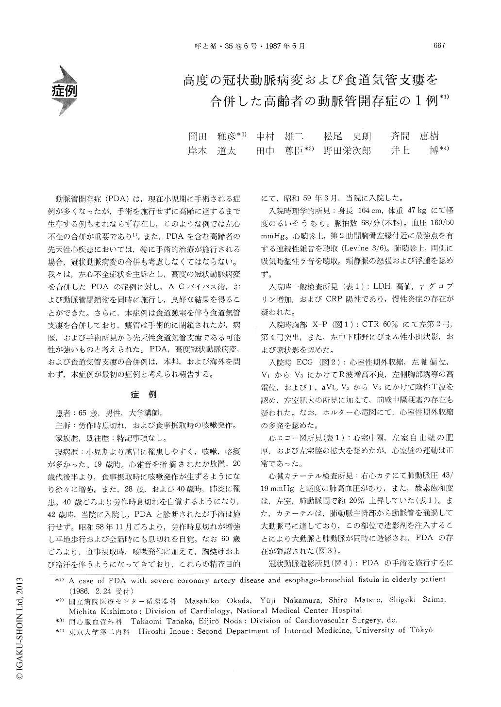
動脈管開存症(PDA)は,現在小児期に手術される症例が多くなったが,手術を施行せずに高齢に達するまで生存する例もまれならず存在し,このような例では左心不全の合併が重要であり1),また,PDAを含む高齢者の先天性心疾患においては,特に手術的治療が施行される場合,冠状動脈病変の合併も考慮しなくてはならない。我々は,左心不全症状を主訴とし,高度の冠状動脈病変を合併したPDAの症例に対し,A-Cバイパス術,および動脈管閉鎖術を同時に施行し,良好な結果を得ることができた。さらに,本症例は食道憩室を伴う食道気管支瘻を合併しており,瘻管は手術的に閉鎖されたが,病歴,および手術所見から先天性食道気管支瘻である可能性が強いものと考えられた。PDA,高度冠状動脈病変,および食道気管支瘻の合併例は,本邦,および海外を問わず,本症例が最初の症例と考えられ報告する。
A 65-year-old man was admitted to the hospital because of exertional dyspnea and paroxysms of coughing after meals. He had been suffering from cough and sputum since his early childhood and had a history of two episodes of pneumonia at the age of 28 and 40 years. He started complaining of exer-tional dyspnea at the age of 40 years and was diag-nosed as having PDA 2 years later. The symptoms became worse several months prior to the admission. Physical examination revealed a grade 3/6 contin-uous murmur at the left upper sternal border and moist rales over the bilateral lower lung fields. Chest roentgenogram showed enlarged cardiac sil-hoette and diffuse shadow in the right lower lung field. Electrocardiogram was significant for the vent-ricular hypertrophy and the suspicious findings of the old anteroseptal myocardial infarction. Right sided catheterization demonstrated mild pulmonary hypertension. The presence of PDA was confirmed by the aortography. Coronary arteriography disclo-sed 90% narrowing in the left main trunk and 75% in both the circumflex coronary artery and the po-sterolateral branch of right coronary artery. Barium swallow showed a fistula connecting the saccular esophageal diverticula and the right bronchus with its orifice identified in the right Bsc bronchus by simultaneous esophago-bronchography. Transbrochial lung biopsy as well as brushing cytological exami-nation gave no definitive evidence of malignancy. Surgical correction was first carried out with respect to the cardiovascular lesions. The operative proce-dures were as follows. Following a median sterno-tomy and the institution of cardiopulmonary bypass, LAD aolta-coronary bypass grafting was performed and then ductus was closed by ligation with 1-0silk and tape. Three weeks later, esophago-bronchial fistula was closed surgically with ligature of 1-0 silk. Around the fistula was there neither inflam-mation nor adhesion of lymph nodes. The post ope-rative course was uneventful and the patient was asymptomatic as of 14 months later.
The implication of this case is that we should consider the possibility of accompanied atheroscle-rotic cardiovascular disease in the elderly patients with congenital heart disease such as PDA, espe-cially when the operative procedure is to be applied to the congenital heart disease. Furthermore this particular case had an esophago-bronchial fistula with esophageal diverticula, etiology of which was most likely of congenital origin according to the history and operative findings.

Copyright © 1987, Igaku-Shoin Ltd. All rights reserved.


