Japanese
English
- 有料閲覧
- Abstract 文献概要
- 1ページ目 Look Inside
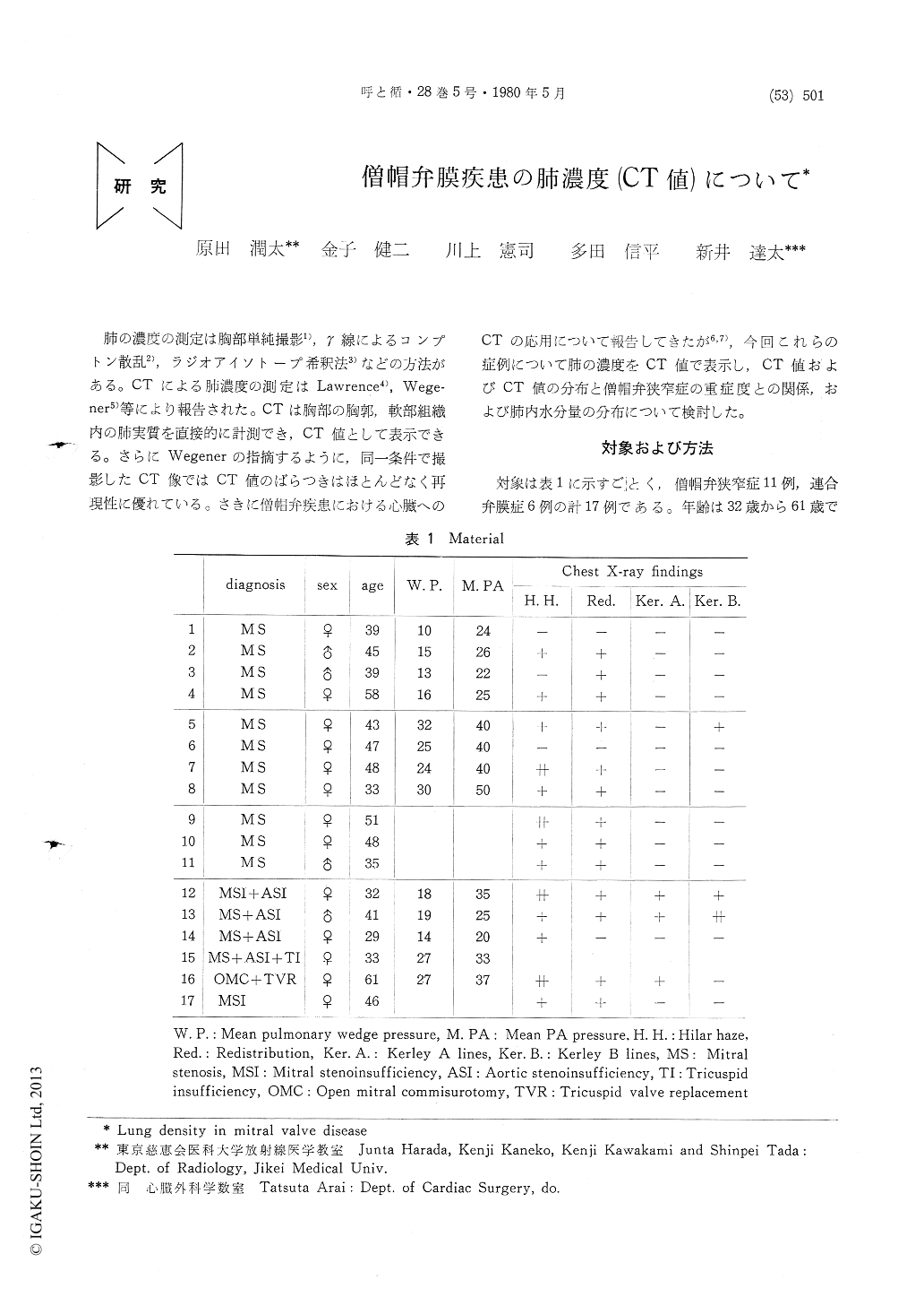
肺の濃度の測定は胸部単純撮影1),γ線によるコンプトン散乱2),ラジオアイソトープ希釈法3)などの方法がある。CTによる肺濃度の測定はLawrence4),Wege—ner5)等により報告された。CTは胸部の胸郭,軟部組織内の肺実質を直接的に計測でき,CT値として表示できる。さらにWegenerの指摘するように,同一条件で撮影したCT像ではCT値のばらつきはほとんどなく再現性に優れている。さきに僧帽弁疾患における心臓へのCTの応用について報告してきたが6,7),今回これらの症例について肺の濃度をCT値で表示し,CT値およびCT値の分布と僧帽弁狭窄症の重症度との関係,および肺内水分量の分布について検討した。
Computed tomography was carried out in 17 patients with cardiac valvular disease for measurement of lung density. In the normal lung, pulmonary density is ranging from -920H to -800H. The distribution of lung density is denser posteriorly than anteriorly and more so in the lower lung.
In severe mitral stenosis (MS), pulmonary density is ranging from -830H to -610H. The distribution of pulmonary density is different and variable in mild MS and combined MS, without evidence of certain pattern. Pulmonary density is greater posteriorly than anteriorly and more so in lower lung in severe MS. The density is not necessarily high in lower lung with Kerley B lines.

Copyright © 1980, Igaku-Shoin Ltd. All rights reserved.


