Japanese
English
- 有料閲覧
- Abstract 文献概要
- 1ページ目 Look Inside
- 参考文献 Reference
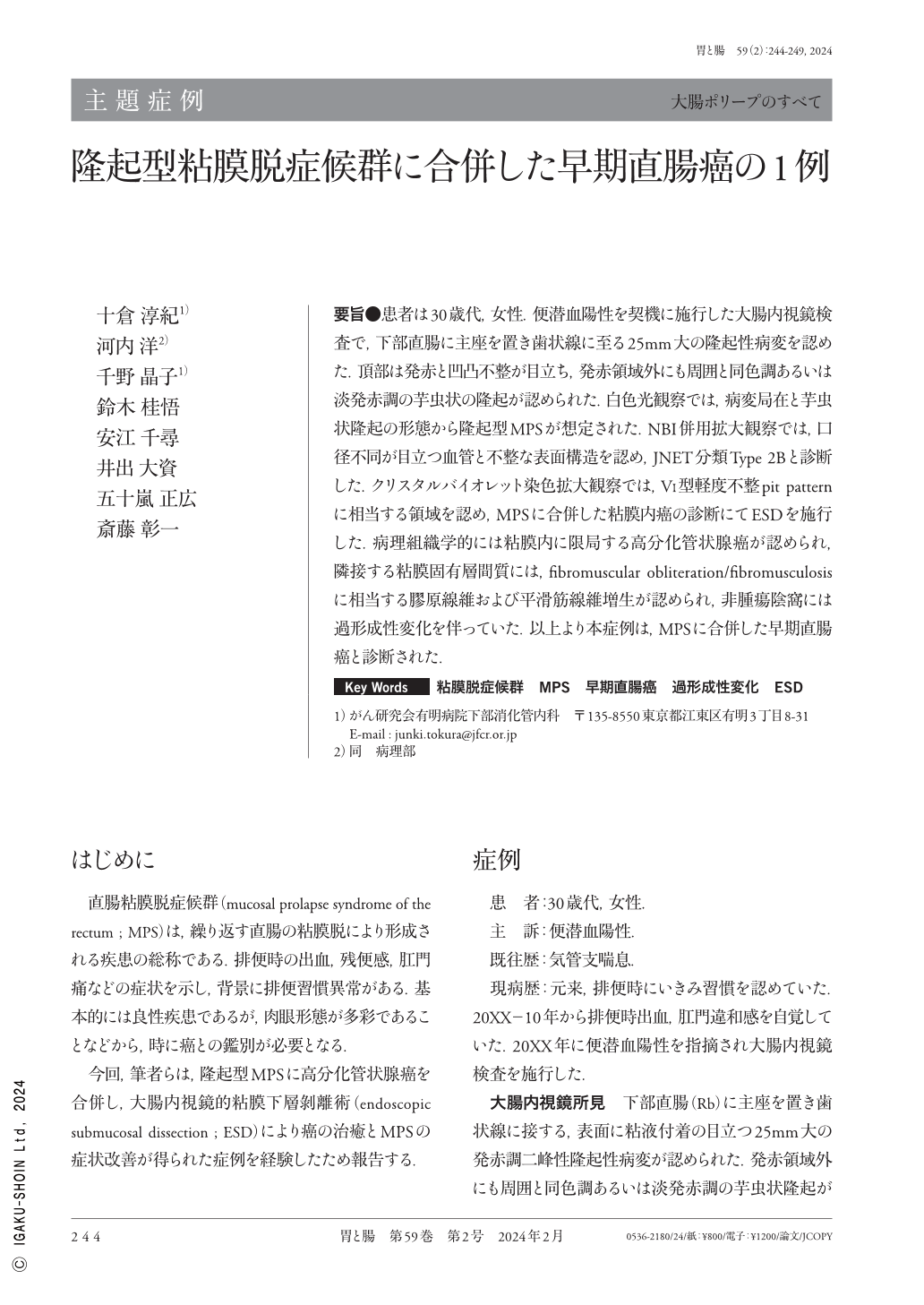
要旨●患者は30歳代,女性.便潜血陽性を契機に施行した大腸内視鏡検査で,下部直腸に主座を置き歯状線に至る25mm大の隆起性病変を認めた.頂部は発赤と凹凸不整が目立ち,発赤領域外にも周囲と同色調あるいは淡発赤調の芋虫状の隆起が認められた.白色光観察では,病変局在と芋虫状隆起の形態から隆起型MPSが想定された.NBI併用拡大観察では,口径不同が目立つ血管と不整な表面構造を認め,JNET分類Type 2Bと診断した.クリスタルバイオレット染色拡大観察では,VI型軽度不整pit patternに相当する領域を認め,MPSに合併した粘膜内癌の診断にてESDを施行した.病理組織学的には粘膜内に限局する高分化管状腺癌が認められ,隣接する粘膜固有層間質には,fibromuscular obliteration/fibromusculosisに相当する膠原線維および平滑筋線維増生が認められ,非腫瘍陰窩には過形成性変化を伴っていた.以上より本症例は,MPSに合併した早期直腸癌と診断された.
A female in her 30s underwent a colonoscopy following a positive fecal occult blood test, which revealed a 25-mm elevated lesion that extended to the dentate line in the lower rectum. The apex of this lesion demonstrated a reddish and irregular appearance, characterizing a caterpillar-like protuberance with the same color tone or displaying a pale erythematous tone similar to the surrounding area that was also observed outside the reddish area. White-light images indicated an elevated MPS(mucosal prolapse syndrome)based on lesion localization and caterpillar-like protuberance morphology. Magnifying endoscopy with NBI(narrow-band imaging)revealed irregular vessels and an uneven surface structure, indicating Japan NBI Expert Team classification Type 2B. Crystal violet staining revealed an area corresponding to Vi mild. Endoscopic submucosal dissection was performed after MPS-associated intramucosal carcinoma diagnosis. Histopathological examination demonstrated a well-differentiated adenocarcinoma confined to the intramucosal layer, accompanied by findings indicating fibromuscular obliteration/fibromusculosis in the adjoining stroma of the intramucosal layer. Based on the results, this case was conclusively diagnosed as MPS-associated early rectal cancer.

Copyright © 2024, Igaku-Shoin Ltd. All rights reserved.


