Japanese
English
- 有料閲覧
- Abstract 文献概要
- 1ページ目 Look Inside
- 参考文献 Reference
- サイト内被引用 Cited by
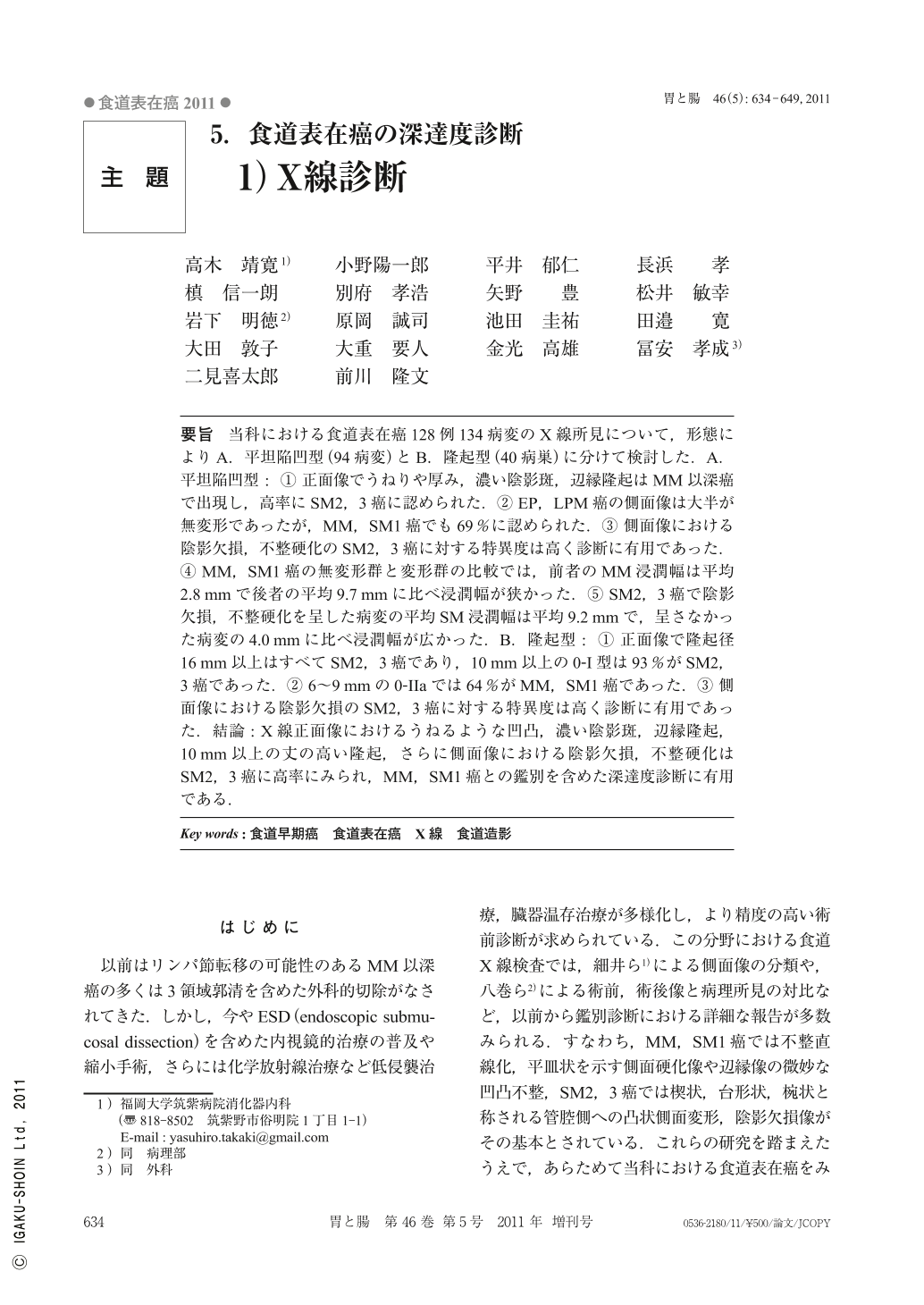
要旨 当科における食道表在癌128例134病変のX線所見について,形態によりA.平坦陥凹型(94病変)とB.隆起型(40病巣)に分けて検討した.A.平坦陥凹型:(1)正面像でうねりや厚み,濃い陰影斑,辺縁隆起はMM以深癌で出現し,高率にSM2,3癌に認められた.(2)EP,LPM癌の側面像は大半が無変形であったが,MM,SM1癌でも69%に認められた.(3)側面像における陰影欠損,不整硬化のSM2,3癌に対する特異度は高く診断に有用であった.(4)MM,SM1癌の無変形群と変形群の比較では,前者のMM浸潤幅は平均2.8mmで後者の平均9.7mmに比べ浸潤幅が狭かった.(5)SM2,3癌で陰影欠損,不整硬化を呈した病変の平均SM浸潤幅は平均9.2mmで,呈さなかった病変の4.0mmに比べ浸潤幅が広かった.B.隆起型:(1)正面像で隆起径16mm以上はすべてSM2,3癌であり,10mm以上の0-I型は93%がSM2,3癌であった.(2)6~9mmの0-IIaでは64%がMM,SM1癌であった.(3)側面像における陰影欠損のSM2,3癌に対する特異度は高く診断に有用であった.結論:X線正面像におけるうねるような凹凸,濃い陰影斑,辺縁隆起,10mm以上の丈の高い隆起,さらに側面像における陰影欠損,不整硬化はSM2,3癌に高率にみられ,MM,SM1癌との鑑別を含めた深達度診断に有用である.
In order to clarify the accuracy of X-ray diagnosis of the depth of invasion, we studied consecutive 134 lesions from 124 cases of superficial esophageal carcinomas. Subjects were divided into the two categories which showed flat or depressed type(A : 94 lesions)and protruding type(B : 40 lesions). The results of flat or depressed type(A)were as follows ; ① In the en-face view, deeper than MM ca showed marked uneven image, dense barium fleck and marginal elevation, and these findings accounted for a high specificity for SM2, 3 ca. ② Most of EP, LPM ca showed lateral image without deformity, and these findings accounted high specificity(69%)for MM, SM1 ca. ③ In the lateral view, the specificity of the finding of marginal deformity and irregular rigidity was high(95%)for SM2, 3 ca. ④ The average width of pathological MM invasion of MM, SM1 ca with non-lateral deformity group was significantly smaller than that with lateral deformity group(2.8mm vs 9.7mm). ⑤ The average width of pathological SM invasion of SM2, 3 ca with marginal defect or irregular rigidity group was significantly larger than that without marginal defect or irregular rigidity group(9.2mm vs 4.0mm). The results of protruding type(B)were as follows ; ① In the en-face view, all the protrusions larger than 16mm in diameter and 93%of gross type 0-I which is larger than 10mm in diameter were SM2, 3 ca. ③ In the lateral view, the specificity of the finding of marginal defect was also high(95%)for protruding SM2, 3 ca. In conclusions ; The en-face view of marked uneven image, dense barium fleck, marginal elevation and gross type 0-I protrusion larger than 10mm in diameter, and in the lateral view of marginal defect and irregular rigidity were useful markers for discriminating SM2, 3 ca from MM, SM1 ca. If these specific radiographic findings are combined to endoscopic findings, indication of endoscopic resection will be much more reliable.

Copyright © 2011, Igaku-Shoin Ltd. All rights reserved.


