Japanese
English
- 有料閲覧
- Abstract 文献概要
- 1ページ目 Look Inside
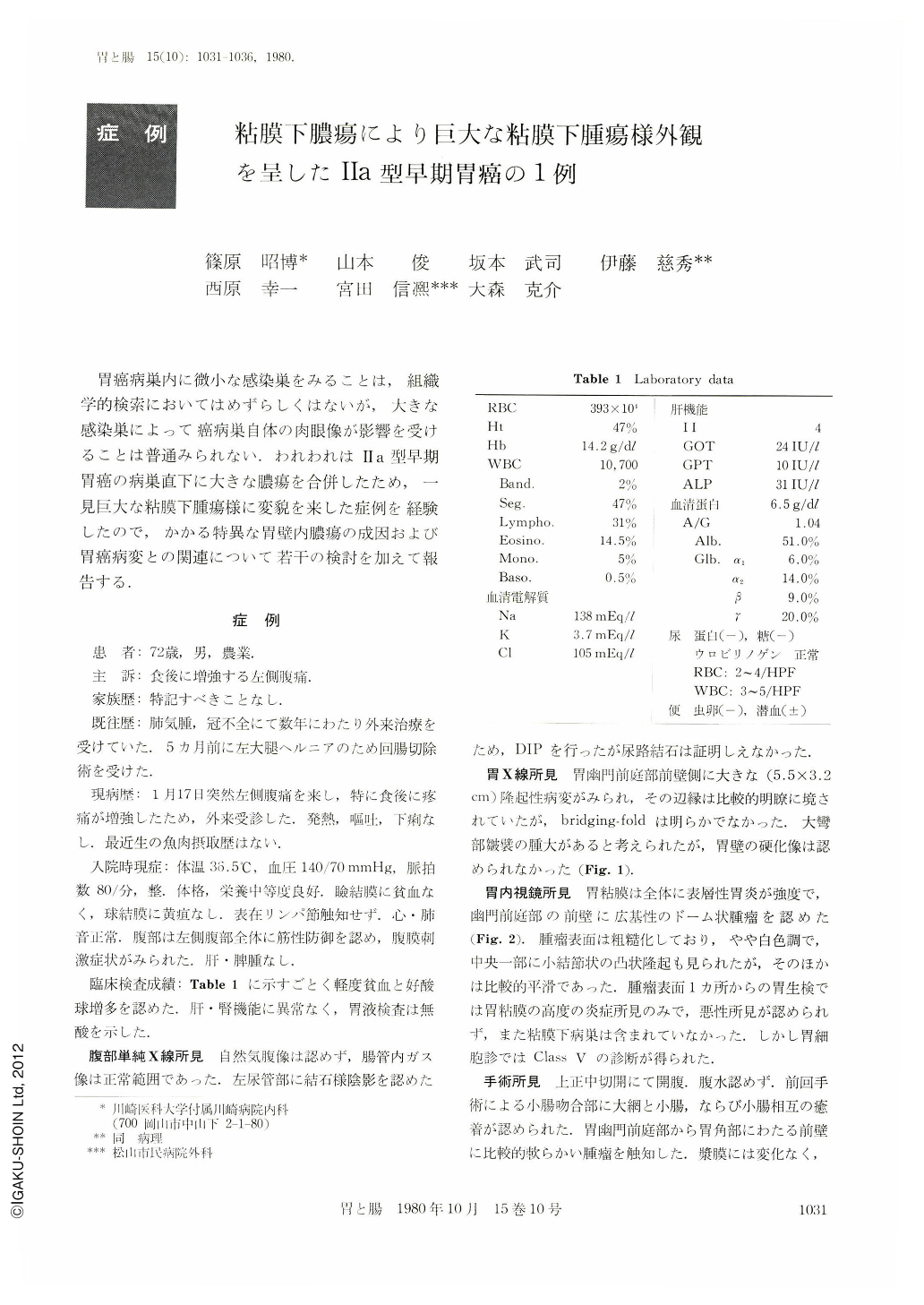
胃癌病巣内に微小な感染巣をみることは,組織学的検索においてはめずらしくはないが,大きな感染巣によって癌病巣自体の肉眼像が影響を受けることは普通みられない.われわれはⅡa型早期胃癌の病巣直下に大きな膿瘍を合併したため,一見巨大な粘膜下腫瘍様に変貌を来した症例を経験したので,かかる特異な胃壁内膿瘍の成因および胃癌病変との関連について若干の検討を加えて報告する.
This was a 72 year-old man patient complaining of sudden abdominal pain and showing muscle guarding over the left half of the abdomen as well as remarkable blood eosinophilia on examination. X-ray study disclosed a large protuberant lesion measuring 5.5 cm in maximum diameter on the anterior wall of the gastric pyloric antrum. Endoscopically the lesion was found sessile, dome-shaped and highly protuberant, showing smooth mucosal surface except for a central small portion of roughening and thus it was suspected to be submucosal tumor. Cytology demonstrated, however, Class V malignancy and therefore, gastrectomy was performed in consideration of advanced cancer.
Grossly the lesion was a hemispherical tumor measuring 3 cm in diameter which was accompanied by definitive bridging folds. On section the submucosal lesion attributable to the protuberant gross appearance was found to consist of a localized discoid focus of chronic abscess measuring 2.6 cm in maximum dimension, which was surrounded by eosinophilic granulation tissues. The mucosa covering the tumor appeared slightly hypertrophic in general and its central small portion was invaded by a solid lesion measuring 1.5×1.0 cm in size extending down to the upper portion of the submucosal abscess. Histologically the covering mucosa was invaded by extremely well-differentiated tubular adenocarcinoma except for its central lesion which consisted of papillary adenocarcinoma. Subsequently, in so far as the malignant portion of this lesion was concerned, it seemed comparable to Ⅱa-type early gastric cancer. A few calcified and lamellar bodies suggestive of parasitic debris were scattered within the submucosal abscess. Although skin test for anisakiasis was pseudopositive at the 30th day after surgery, the blood eosinophilia disappeared completely at that time and thus the submucosal abscess seemed most likely to be parasitic, particularly of anisakiasis.
Only a few cases of compound gastric cancer and anisakiasis have been reported in Japan, so that definite relation between both lesions remains obscure. In the present study, however it is assumed that gastric carcinoma lesions comprising well-differentiated adenocarcinomas could possibly facilitate the migration of anisakis larva there even when they be non-ulcerated. A few available parameters are presented for preoperative evaluation on the macroscopic modification of early gastric cancer by submucosal abscesses.
Copyright © 1980, Igaku-Shoin Ltd. All rights reserved.