Japanese
English
- 有料閲覧
- Abstract 文献概要
- 1ページ目 Look Inside
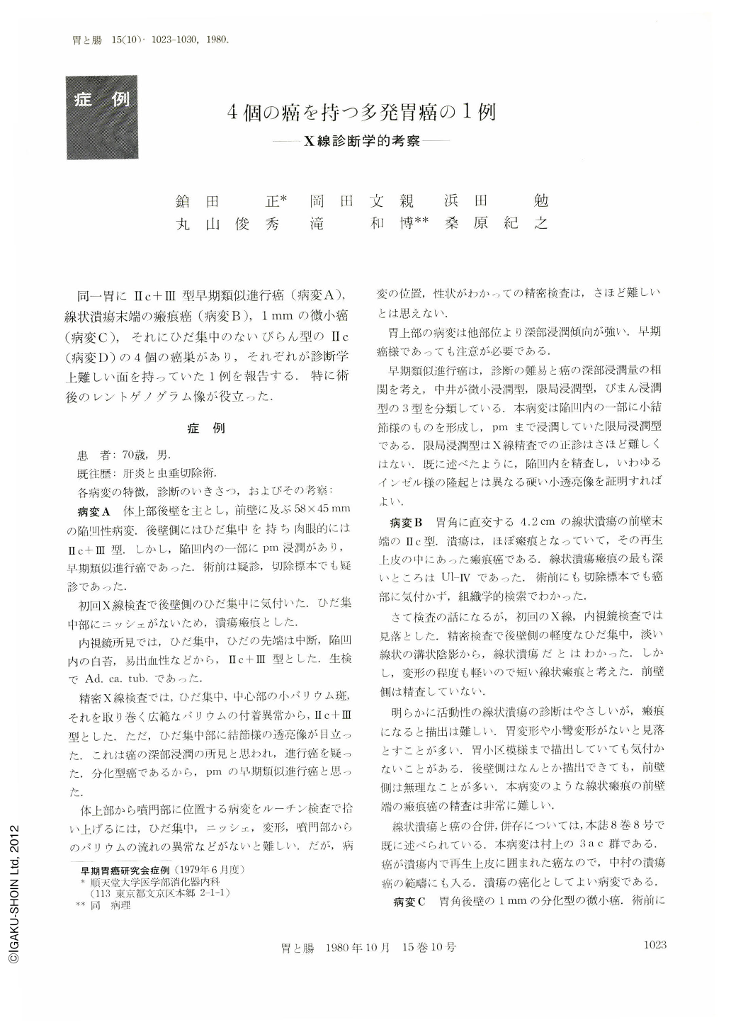
同一胃にⅡc+Ⅲ型早期類似進行癌(病変A),線状潰瘍末端の瘢痕癌(病変B),1mmの微小癌(病変C),それにひだ集中のないびらん型のⅡc(病変D)の4個の癌巣があり,それぞれが診断学上難しい面を持っていた1例を報告する.特に術後のレントゲノグラム像が役立った.
The resected stomach of 70 year-old man was radiologically (postoperative roentgenogram), macroscopically and histologically carefully examined, and then 4 foci of gastric cancer, Ⅱc+Ⅲ like advanced cancer (Lesion “A”), scar-carcinoma coexistent with linear ulcer near its end (Lesion “B”), microcarcinoma about 1 mm in diameter (Lesion “C”) and Ⅱc type early gastric cancer (Lesion “D”), were found. From diagnostic point of view, there remain several problems awaiting to be solved in each lesion, and diagnostic information was displayed on the postoperative roentgenogram.
1. The reported incidence of multiple gastric cancers with more than 4 foci was between 0.13 (Kitaoka et al) and 0.58% (Koba et al), and 0.24% of our materials were found to have more than 4 foci.
2. Lesion “A”, Ⅱc+Ⅲ like advanced cancer was preoperatively correctly diagnosed. Lesion “D”, Ⅱc type early gastric cancer was macroscopically noted in the resected specimen, and the remaining lesions were only found by the histological examination.
3. Lesion “A” and “D” were so clearly visualized on the postoperative roentgenogram that an accurate diagnosis was made. Lesion “B” and “C” were thought as benignancy on the postoperative roentgenogram.
4. For an accurate x-ray diagnosis of localized advanced cancer which apparently seems to be depressed type early cancer, it is necessary to well examine the bottom of depression and to document a rather solid elevated lesion different from socalled “Island”.
5. It seems to be very difficult to make an x-ray diagnosis of ulcer-carcinoma near the end of linear ulcer, and gastric biopsy is indispensable to establish a diagnosis.
6. Lesion “B” belongs to 3 a c group of Murakami's classification of ulcer-carcinoma and it also meets the qualification for ulcer-carcinoma by Nakamura.
7. Microcarcinoma around 1 mm in diameter, if it shows depression, can be visualized on the postoperative roentgenogram.
8. Lesion “D” was visualized as type Ⅱc early gastric cancer on the postoperative roentgenogram, but the histological examination of its serial sections is suggestive of cancer development in the focus of atypical epithelial lesion.

Copyright © 1980, Igaku-Shoin Ltd. All rights reserved.


