Japanese
English
- 有料閲覧
- Abstract 文献概要
- 1ページ目 Look Inside
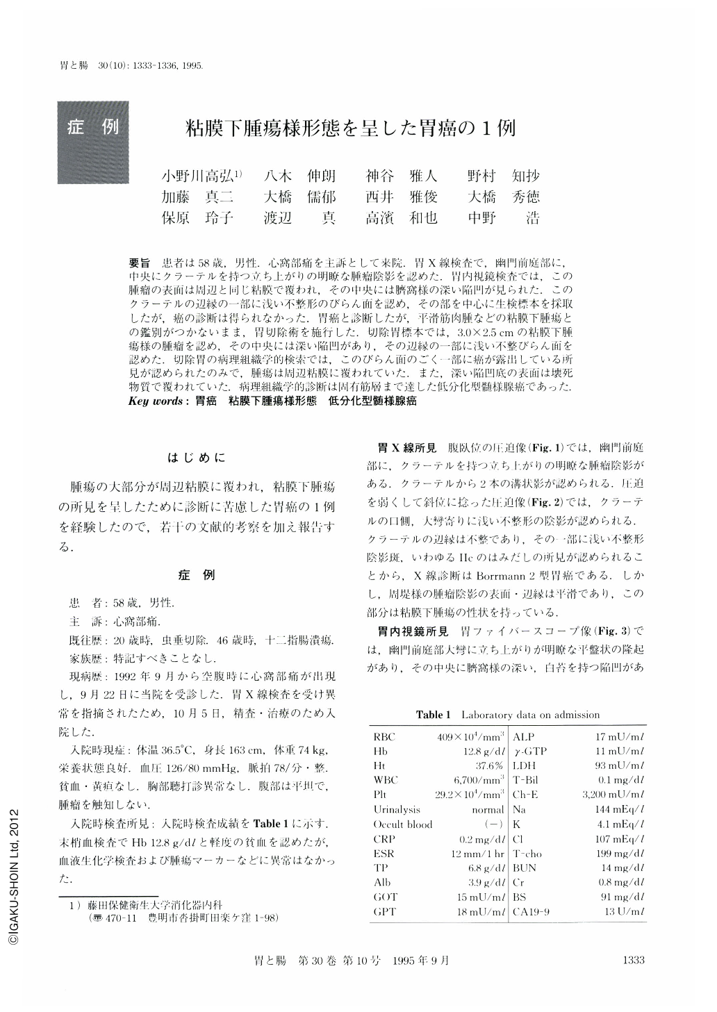
要旨 患者は58歳,男性.心窩部痛を主訴として来院.胃X線検査で,幽門前庭部に,中央にクラーテルを持つ立ち上がりの明瞭な腫瘤陰影を認めた.胃内視鏡検査では,この腫瘤の表面は周辺と同じ粘膜で覆われ,その中央には臍窩様の深い陥凹が見られた.このクラーテルの辺縁の一部に浅い不整形のびらん面を認め,その部を中心に生検標本を採取したが,癌の診断は得られなかった.胃癌と診断したが,平滑筋肉腫などの粘膜下腫瘍との鑑別がつかないまま,胃切除術を施行した.切除胃標本では,3.0×2.5cmの粘膜下腫瘍様の腫瘤を認め,その中央には深い陥凹があり,その辺縁の一部に浅い不整びらん面を認めた.切除胃の病理組織学的検索では,このびらん面のごく一部に癌が露出している所見が認められたのみで,腫瘍は周辺粘膜に覆われていた.また,深い陥凹底の表面は壊死物質で覆われていた.病理組織学的診断は固有筋層まで達した低分化型髄様腺癌であった.
A 58-year-old man was admitted to our hospital with the complaint of epigastric pain. In the radiological examination, a tumor shadow with a large crater was demonstrated in the antrum. At the edge of the crater, a shallow depression was also seen. Endoscopic pictures showed a Submucosal tumor-like mass and the central deep depression. The shallow, erosive depression was Seen at the edge of the deep depression. Cancer cells were not revealed in the biopsy specimen taken from this shallow depression and at the edge of the deep depression. However, the possibility of a malignant tumor such as an atypical type of gastric cancer or leiomyosarcoma was not able to be excluded. Gastrectomy was performed. In the resected specimen, Submucosal tumor-like mass, measuring 3.0 X 2.5 cm, with deep depression was found in the anterior wall of the antrum. At the edge of the deep depression, Ⅱc-like shallow depression was seen. Histologically, the tumor was covered with surrounding mucosa and cancer cells were revealed only in the small part of the shallow depression. The base of the deep part of the depression was covered with necrotic tissue. The type of cancer was poorly differentiated adenocarcinoma, medullary type, and the invasion of the cancer was limited to the proper muscle layer. In this case, the correct diagnosis was not easy to reach because of the cancer mass being almost covered with the surrounding mucosa.

Copyright © 1995, Igaku-Shoin Ltd. All rights reserved.


