Japanese
English
- 有料閲覧
- Abstract 文献概要
- 1ページ目 Look Inside
Barret's esophagusと呼ばれる病変は下部食道粘膜が円柱上皮に置換された状態をさし,Barrett(1950)の報告にその名を由来する.
1957年以降,このBarrett's esophagusが腺癌の発生母地として注目されてきた.われわれは剖検例ではあるがBarrett's esophagusに腺癌を合併した症例を経験した.本邦の報告例は調査した範囲内では学会発表例以外見当たらない.最近の進歩したX線造影,内視鏡観察の普及からみて,臨床上かかる例に遭遇する機会も少なくないと思われるので,病理学的所見を中心に記載する.
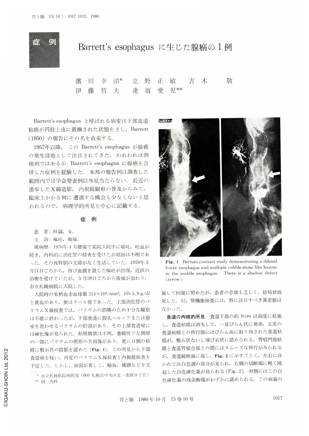
An 81 year-old woman was admitted to Sapporo City General Hospital for intermittent hematemesis, vomiting and abdominal pain. Examination of the peripheral blood revealed anemia showing 5.9 g/100 ml of hemoglobin and 214×104/mm3 of red blood cells. X-ray examination of the upper digestive tract by barium meal demonstrated a dilated esophagus with coarse mucosal surface intermixed with cobble-stone appearance, and a shadow defect. The stomach appeared normal. Esophageal carcinoma was suspected. The patient, however, emaciated insidiously. The complete examination was not carried out before the patient's death. At autopsy, the lower esophagus was dilated and replaced by reddish mucosa with shallow ulcer, which smoothly shifted to the fundic mucosa. Two slightly elevated indurations with whitish gray color were found in the left lower wall surrounded by coarse mucosa. Thickened esophageal mucosa, showing cobble-stone appearance, was also noted in the ulcer, which was located in the border between the lower and middle esophagus. Histologically, the reddish mucosa was similar to the fundic mucosa of the stomach (Barrett's esophagus), though atrophic and accompanied with lymphoid cell infiltration. The indurated lesions in the left wall were distinct tubular adenocarcinoma infiltrating into the submucosa. The coarse mucosa surrounding the induration showed a histologic appearance of in situ carcinoma.

Copyright © 1980, Igaku-Shoin Ltd. All rights reserved.


