Japanese
English
- 有料閲覧
- Abstract 文献概要
- 1ページ目 Look Inside
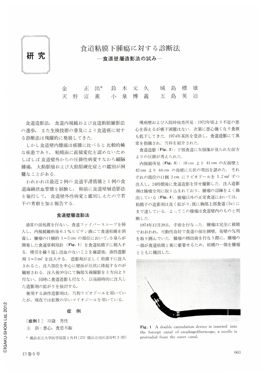
食道造影法,食道内視鏡および食道動脈撮影法の進歩,また生検技術の普及により食道癌に対する診断法は飛躍的に発展してきた.
しかし食道壁内腫瘍は癌腫に比べると比較的稀な疾患であり,粘膜面に直接変化を認めないためしばしば食道壁外からの圧排性病変すなわち縦隔腫瘍,大動脈瘤および大動脈硬化症との鑑別が困難なことがある.
Recently diagnostic methods for esophagus cancer have made a remarkable progress, because esophagogram, esophagoendoscopy and esophagoarteriography have been improved, and biopsy techniques have also been coming into wider use.
However, intramural tumor of the esophagus is a relatively rare disease. As it shows no changes in the mucosal surface, it is often difficult to differentiate it from extrinsic lesions out of the esophagus, namely, mediastinal tumor, aortic aneurysm, and sclerosis of aorta.
We have recently encountered two cases of esophagoleiomyoma and a case of cavernous hemangioma of the esophagus, for which we performed intramural esophagography.
As a result, we were able to differentiate these lesions from extrinsic one of the esophagus. In this method, under esophagofiberscope, a double cannula is inserted into the biopsy canal of the esophagofiberscope and oiled contrast medium is injected in the submucosal layer few centimeter oral to the tumor. When contrast medium is injected correctly, a roundshaped protrusion around the injected site is observed. After this procedure, chest X-ray and routine esophagogram is taken under different positions to observe spreading of contrast medium. The contrast medium injected by this method spreads in the submucosal layer and seeps through out of the muscle layer forming, as it were, two layers, submucosal and extramural, and encloses intramural tumor. This method we have devised appears to make definite the diagnosis of intramural tumor of the esophagus.
This method is named 〔Intramural Esophagography〕 and we are going to make further investigation.

Copyright © 1977, Igaku-Shoin Ltd. All rights reserved.


