Japanese
English
- 有料閲覧
- Abstract 文献概要
- 1ページ目 Look Inside
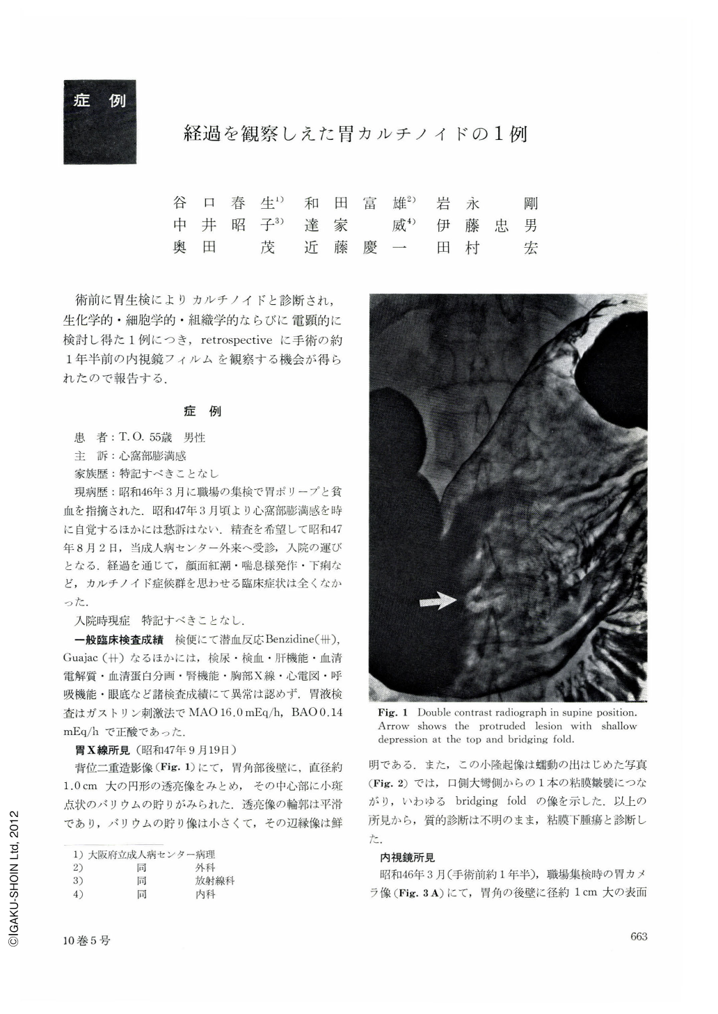
術前に胃生検によりカルチノイドと診断され,生化学的・細胞学的・組織学的ならびに電顕的に検討し得た1例につき,retrospectiveに手術の約1年半前の内視鏡フィルムを観察する機会が得られたので報告する.
One and half a year before a 55-year-old patient had been suspected to have a gastric polyp. When he was reexamined by x-ray and endoscopy, a tentative diagnosis was submucosal tumor of the stomach. This in turn was determined to carcinoid after biopsy. During the course of follow-up the shape and size of the tumor remained unaltered. Biochemical examinations including measurement of 5-HT, 5-HIAA, gastrin, histamine and catecholamine were performed. Furthermore, blood was taken during operation separately from the artery and vein in the tumor region to compare the levels of 5-HT in each of blood samples, but they all turned out to be within normal range. The small size of the tumor and substances selected for measurement probably accounted for this result. The tumor was gastric carcinoid in early stage, measuring only 7 mm in diameter, mostly developing in the submucosa of the posterior wall of the lower part of the body. Histologically it had a typical structure consisting of solid cell nests. Argyrophil granules were demonstrated in the cytoplasm. Electron microscopy also revealed small rounded secretory granules. Optical microscopy further showed vesiculation in the cytoplasm, corresponding to vesicular mitochondria noticed by electron microscopy. In the nuclear chromatin as well this was recognized as rough agglutination, a feature to be seen by both types of microscope.

Copyright © 1975, Igaku-Shoin Ltd. All rights reserved.


