Japanese
English
- 有料閲覧
- Abstract 文献概要
- 1ページ目 Look Inside
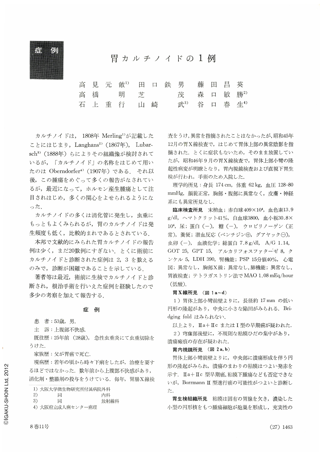
カルチノイドは,1808年Merling1)が記載したことにはじまり,Langhans2)(1867年),Lubarsch3)(1888年)らによりその組織像が検討されているが,「カルチノイド」の名称をはじめて用いたのはOberndorfer4)(1907年)である.それ以後,この腫瘍をめぐって多くの報告がなされているが,最近になって,ホルモン産生腫瘍として注目されはじめ,多くの関心をよせられるようになった.
カルチノイドの多くは消化管に発生し,虫垂にもっともよくみられるが,胃のカルチノイドは発生頻度も低く,比較的まれであるとされている.
Carcinoid tumor of the stomach is a relatively rare disease. The case presented here comes to be the twenty-first in Japan, and is the last following three reported cases which were preoperatively diagnosed as gastric carcinoid.
The patient was a 54-year-old man whose chief complaints were abdominal distress.
X-ray and the endoscopic examinations of the stomach revealed a small circumscribed, protruding lesion with a central depression on the anterior wall of the mid-body.
The lesion was diagnosed as carcinoid tumor on the study of biopsy.
The gross specimen of totally resected stomach showed a round, elevated tumorous lesion, 17×13×5 mm, with a central ulceration on the anterior wall of the mid-body near the lesser curvature, and other two ulcer scars in the fundus.
Histopathologically carcinoid was confirmed with the tumor mass which was largely located in the submucosal layer, though partly infiltrating into the serosa. Argyrophil reaction by Bodian's method was positive for tumor cells, and electron microscopy disclosed intracytoplasmic secretory granules within tumor cell bodies. There were metastatic lymph nodes along the lesser curvature, but no metastasis in the liver.
None of carcinoid syndromes was shown in the patient, and Serotonin level in the blood as well as urinary 5-HIAA remained within normal values.
The lesions on the fundus were found to be lymphoreticular hyperplasia on microscopy.
The patient is alive and well twenty four months after the operation.

Copyright © 1973, Igaku-Shoin Ltd. All rights reserved.


