Japanese
English
- 有料閲覧
PPV購入案内
1,320円 (1,200円+税10%) こちらは、795ページから801ページの文献です。購入には医書.jp本会員登録が必要です。
会員登録完了後に改めて上記「購入サイトへ」を選択してください。
または「購入サイトへ」選択後に「購入ログイン」、「新規会員登録」の順に進んでください。
会員登録について詳しくはをご確認ください。
- Abstract 文献概要
- 1ページ目 Look Inside
今回,私どもは胃X線,内視鏡検査上興味ある所見を示した胃カルチノイドの1例を経験したので,そのX線,内視鏡所見を中心に報告する.
患 者:加○ 昇 52歳 男性
主 訴:なし(人間ドック)
既往歴・家族歴:特になし.
経 過:昭和46年8月人間ドックで胃に異常所見なし,昭和48年3月再度の人間ドックで胃X線検査で異常所見を指摘され,胃内視鏡,生検の結果,胃癌と診断され昭和48年5月胃切除術をうける.
Abnormal finding in the stomach was pointed out to a 52-year old male patient in the course of human dock examination. He has had no carcinoid symptoms, though urine 5-HIAA and serum serotonin was not checked. The presext paper deals with the radiographic, endoscopic and exoloratory findings of this case.
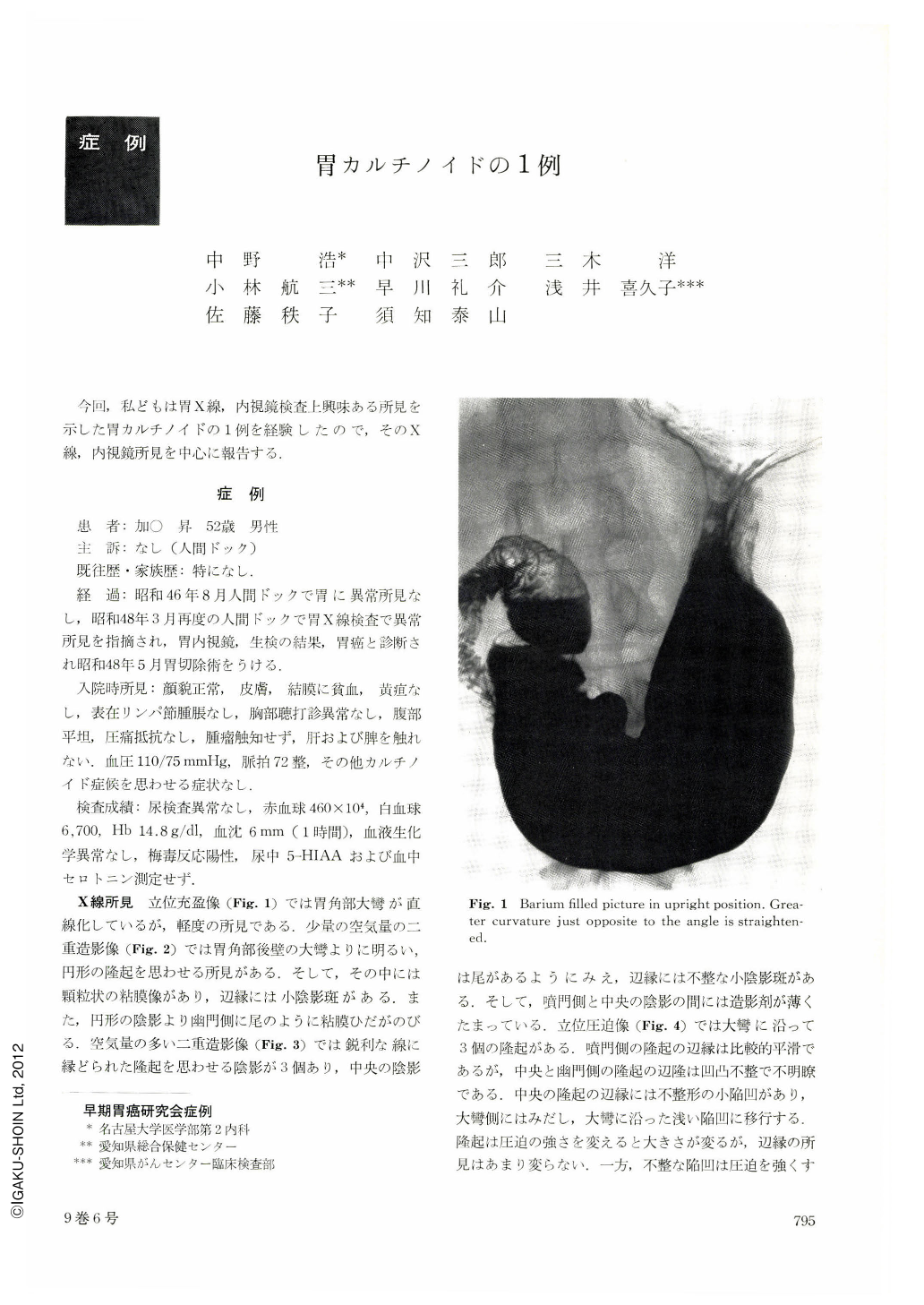
Radiographically we found three protruded lesions, hard to determine whether of epithelial or nonepithelial natnre. On the surface of the lesions and among them, there existed irregular depressions and the radiographical diagnosis was gastric sarcoma or Ⅱc+Ⅱa type. Endoscopy showed findings of multiple submucosal tumors.
The resected specimen showed that there were at the lower gastric corpus an irregularly depressed portion and three submucosal tumors around it. Histological diagnosis was carcinoid confirmed as such after argyrophil staining and electron-microscopy.
Cut specimen showed that tumor remained mainly in the deep Iayer of the mucosa and the location of this tumor was exactly reflected in our x-ray finding. We might call this finding as that of “epithelial submucosal tumor”.
Copyright © 1974, Igaku-Shoin Ltd. All rights reserved.