Japanese
English
- 有料閲覧
- Abstract 文献概要
- 1ページ目 Look Inside
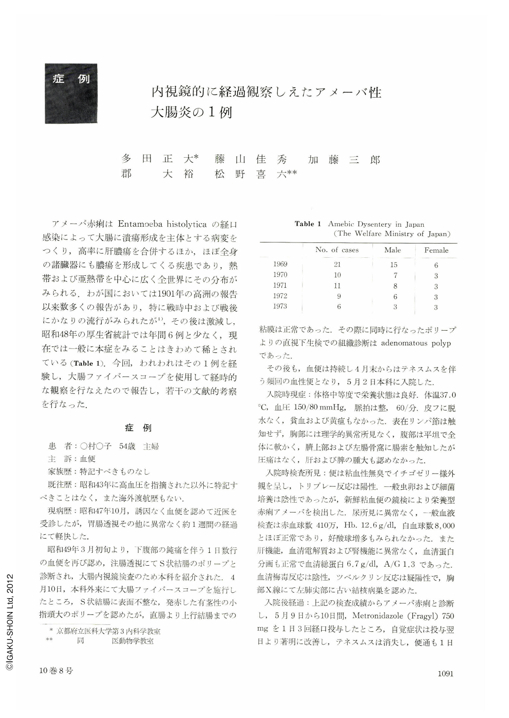
アメーバ赤痢はEntamoeba histolyticaの経口感染によって大腸に潰瘍形成を主体とする病変をつくり,高率に肝膿瘍を合併するほか,ほぼ全身の諸臓器にも膿瘍を形成してくる疾患であり,熱帯および亜熱帯を中心に広く全世界にその分布がみられる.わが国においては1901年の高洲の報告以来数多くの報告があり,特に戦時中および戦後にかなりの流行がみられたが1),その後は激減し,昭和48年の厚生省統計では年間6例と少なく,現在では一般に本症をみることはきわめて稀とされている(Table 1).今回,われわれはその1例を経験し,大腸ファイバースコープを使用して経時的な観察を行なえたので報告し,若干の文献的考察を行なった.
Recently, with the diffusion of sanitary thought and remarkable advances of medicine, amebic dysentery has been rapidly decreasing in Japan. In 1973, only 8 cases of amebic dysentery were reported to Japanese Welfare Ministry.
In this paper, a case of amebic dysentery, diagnosed and followed up by colonofiberscopy, was described, and the endoscopical findings of amebic dysentery was discussed, comparing with those of other colonic inflammatory diseases, especially colitis ulcerosa.
The patient, a 54-year-old woman, suffered from bloody diarrhea and tenesmus, and she was admitted to our University hospital. Five days after the onset, the first endoscopy was performed, and multiple scattered aphta-like small erosions or shallow ulcers were observed on the rectal mucosa, but the intervening mucosa among them showed no gross pathological changes. By dye scattering method, they were clearly and distinctly observed, and on the surface of the intervening mucosa, minute mucosal appearances were clso clearly demonstrated. At the same time, biopsy examination was performed, and Entamoeba histolytica was histologically found in the coats of ulcers and identified as such parasitologically. On the 11 th day, the second endoscopy revealed typical amebic ulcers, 5~7 mm in diameter, with the edematous and inflammatory intervening mucosa at the same region. After the medical treatment, these ulcers and mucosal inflammation disappeared, and only multiple small ulcer scar formations, clearly demonstrated by dye scattering method, remained.

Copyright © 1975, Igaku-Shoin Ltd. All rights reserved.


