Japanese
English
- 有料閲覧
- Abstract 文献概要
- 1ページ目 Look Inside
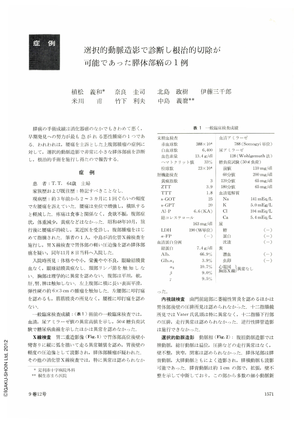
膵癌の手術成績は消化器癌のなかでもきわめて悪く,早期発見への努力が最も急がれる悪性腫瘍の1つである.われわれは,腰痛を主訴とした上腹部腫瘤の症例に対して,選択的動脈造影で非常に小さな膵体部癌を診断し,根治的手術を施行し得たので報告する.
症例
患 者:T. T. 64歳 主婦
家族歴および既往歴:特記すべきことなし.
現病歴:約3年前から2~3カ月に1回ぐらいの頻度で左腰痛を訴えていた,腰痛は坐位で増強し,横臥すると軽減した.とう痛は食事と関係なく,食欲不振,腹部症状,体重減少,黄疸などはなかった.昭和48年10月,旅行後に腰痛が持続し,某近医を受診し,腹部腫瘤をはじめて指摘された.筆者の1人,中島が消化管X線検査を施行し,胃X線検査で胃体部の軽い圧迫像を認め膵体部癌を疑い,同年11月8日当科へ入院した.
The early diagnosis and curative resection of the pancreas cancer is still difficult, in spite of the recent progress in the diagnostic procedures. Using the selective angiography a very small pancreas body cancer as small as 2 cm in diameter could be diagnosed and successfully operated. An old housewife 64 years of age had been suffered from left back pains of three years duration, any abdominal pain, weight loss, or jaundice. An abdominal mass, 6×3 cm in size was palpated in the left upper abdominal quadrant. The patient was transferred to our surgical ward on Nov. 8. 1973.
In the laboratory findings, serum and urine amylase levels were increased. The glucose tolerance test was slightly abnormal. Another findings all were within normal limits. Gastrointestinal series revealed light compression on the posterior wall of the body of the stomach. No abnormal findings was observed by gastroduodenoscopy. Catheterization into the pancreatic duct was impossible at pancreatography, in despite of our carefull efforts. In the selective caeliac angiography, splenic, common hepatic arteries were normal. The dorsal pancreatic artery was obstructed with dilatation and irregular encasement. Abnormal small vessels were visualized in the pancreas body. The configuration of this abnormal vascularization measured rounded about 2 cm in diameter. In the capillary phase, the increased parenchymal accumulation of the contrast medium was not revealed in the mass but in the pancreas tail. In the venous phase, splenic vein was displaced and compressed upward with doom formation and diffuse narrowing. Following the arteriographic findings it may probably be diagnosed as a small pancreas body cancer with chronic pancreatitis. Curative operation was performed on Dec. 8. 1973. In the pancreas body, the grayish white tumor was visible. There was no invasion and no metastasis. Hystological diagnosis was adenocarcinoma tubulare with mucinous cells and scirrhous, other non tumorous region was chronic pancreatitis. Postoperative course was uneventful and discharged on 21 th postoperative day. After the operation, glucose tolerance test was slightly abnormal but soon returned to the normal pattern. Mitomycin C, 5-Fu and cylocide were given as anticancerous chemotherapy for 5 weeks. Nine months after the operation, the patient is in good condition and no sign of recurrence.

Copyright © 1974, Igaku-Shoin Ltd. All rights reserved.


