Japanese
English
- 有料閲覧
- Abstract 文献概要
- 1ページ目 Look Inside
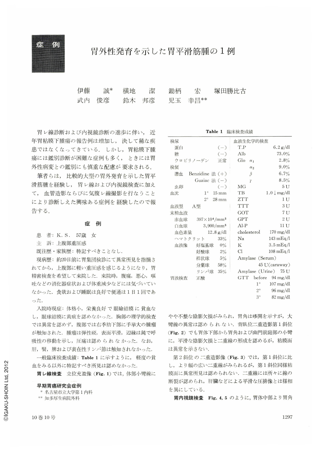
胃レ線診断および内視鏡診断の進歩に伴い,近年胃粘膜下腫瘍の報告例は増加し,決して稀な疾患ではなくなってきている.しかし,胃粘膜下腫瘍には鑑別診断が困難な症例も多く,ときには胃外性病変との鑑別にも慎重な配慮が要求される.
筆者らは,比較的大型の胃外発育を示した胃平滑筋腫を経験し,胃レ線および内視鏡検査に加えて,血管造影ならびに気腹レ線撮影を行なうことにより診断しえた興味ある症例を経験したので報告する.
A fifty-seven-year-old female visited our hospital complaining of upper abdominal discomfort. Physical examination revealed a tumor measuring 10 cm by 5 cm in diameter on the right upper quadrant, which was elastic hard in consistency and movable on respiration.
Roentgenologic study of the stomach showed the double lines from the lower-corpus to the antrum on the lesser curvature, and they were not a series at several points. The double lines varied by the change of patient's position and by respiration, but abnormal findings were not recognized on the mucosa.
Endoscopical study revealed a smooth polypoid lesion with bridging folds from the lower-corpus to the antrum on the lesser curvature.
Pneumoperitoneography and celiac angiography were done to differentiate it from an extra-gastric lesion and to confirm the location, diameter and malignancy of the tumor.
Pneumoperitoneogram showed a tumor shadow corresponding with the lesion on the right upper quadrant. Angiogram revealed an irregular neovascularization over the region of the tumor shadow, demonstrating blood supply by the right gastric and gastroduodenal artery to it. The tumor shadow was about 9 cm by 10 cm in size, but it was difficult to determine whether or not it was malignant.
General impression of this lesion was a submucosal tumor, most likely a leiomyoma of the stomach.
The pathological diagnosis was leiomyoma which showed extragastric growth from the serosal side of the lesser curvature of the lower-corpus.
This case was characterized by a large and extragastric-growth leiomyoma without ulceration and in roentgenological findings by the double lines with their discontinuity.

Copyright © 1975, Igaku-Shoin Ltd. All rights reserved.


