Japanese
English
- 有料閲覧
- Abstract 文献概要
- 1ページ目 Look Inside
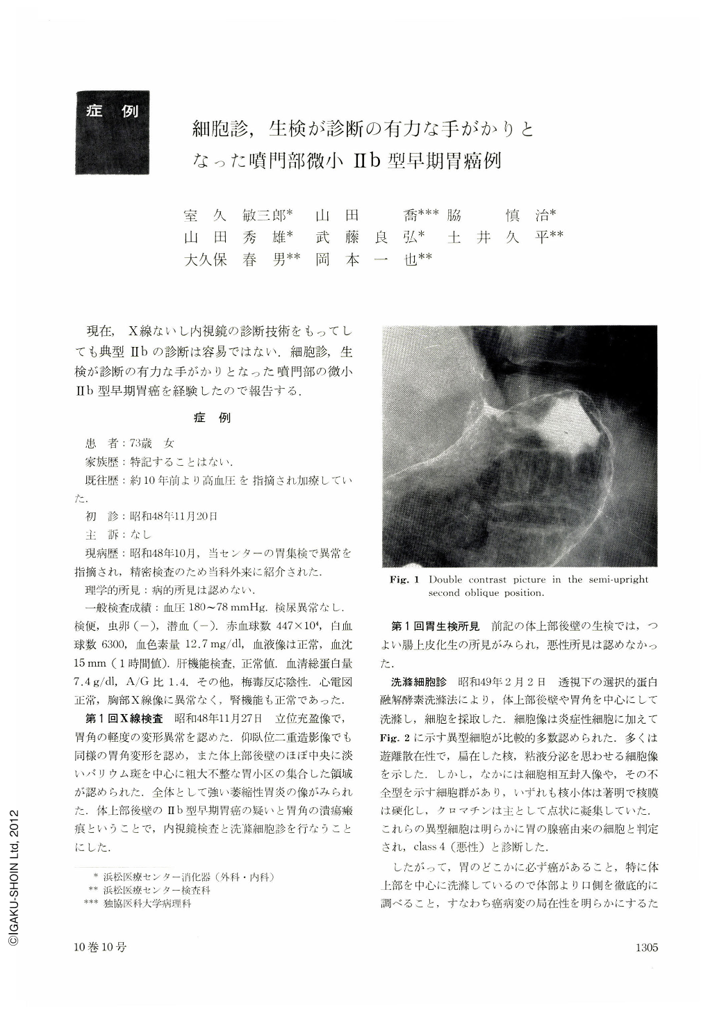
現在,X線ないし内視鏡の診断技術をもってしても典型Ⅱbの診断は容易ではない.細胞診,生検が診断の有力な手がかりとなった噴門部の微小Ⅱb型早期胃癌を経験したので報告する.
The patient, a 73 years old female, had no symptoms to complain of, but an abnormality of the stomach was pointed out at a gastric mass survey. She was referred to our Hospital for further GI. study.
The first upper GI. study showed suspicious early cancer at the posterior wall of the upper body, but no malignancy was proved in both gastrofiberscopic finding and biopsy specimen taken from the cardia and upper body.
The cytologic examination by a selective proteolytic lavage method disclosed malignant cells.
The second gastrofiberscopic biopsy showed early cancer in a piece taken from the cardia; but the difinite site was undetermined.
Gross finding of extirpated stomach in fresh and half-fixed specimens hardly gave hint of cancer lesion, whereas atrophic gastritis with intestinal metaplasia on the gastric mucosa was evident, but the lesion in fixed specimen at the lesser curvature near the posterior wall of the esophago-gastric junction showed slightly more gray in colour than any other surrounding mucosa.
Histologically, the lesion was adenocarcinoma tubulare measuring only 8×4 mm in size and located within the mucosal layer.

Copyright © 1975, Igaku-Shoin Ltd. All rights reserved.


