Japanese
English
- 有料閲覧
- Abstract 文献概要
- 1ページ目 Look Inside
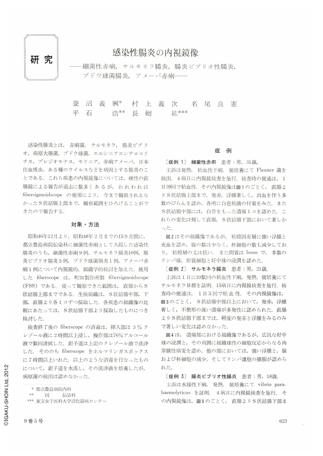
感染性腸炎とは,赤痢菌,サルモネラ,腸炎ビブリオ,病原大腸菌,ブドウ球菌,エルシニアエンテロコリチカ,プレジオモナス,モリニア,赤痢アメーバ,日本住血吸虫,ある種のウイルスなどを病因とする腸炎のことである.これら疾患の内視鏡像については,硬性の直腸鏡による報告が過去に数多くあるが,われわれはfibersigmoidscopeの使用により,今まで観察されえなかったS状結腸上部まで,観察範囲をひろげることができたので報告する.
Among cases of infectious enteritis admitted to the Tokyo Metropolitan Toshima Hospital under a clinical diagnosis of bacillary dysentery, endoscopic and histological studies were carried out in 9 cases of bacillary dysentery, 10 cases of salmonella enteritis, 5 cases of enteritis due to vibrio parahemoliticus, 1 case of staphylococcal enteritis, and 1 case of amoebic dysentery. Fibersigmoidoscope was used to study the range from the rectum to upper sigmoid colon.
As to the endoscopic picture of infectious enteritis in the authors' own experience, edema, reddening, erosion or ulcer constituted the general findings. In the histological picture, edema, hyperemia, erosion or ulcer, and infiltration by inflammatory cells represented the general findings.
In bacillary, dysentery remarkable findings such as reddening, erosion and ulcer were frequently found in the lower part of sigmoid colon. In salmonella enteritis, unlike dysentery, remarkable changes were frequently seen in the middle and upper parts of the sigmoid colon. And in one case, multiple ulcers were noted. In vibrio enteritis, edema was the main finding in all cases, without remarkable findings such as erosion and ulcer. Main sites of change were the rectum and lower part of the sigmoid colon. Histologically ocal accumlations of lymphocytes were seen in 2 lcases. This finding was not seen in others.
While some difference was noted in the severity of the lesion and site of the main lesion, no endoscopic or histological findings capable of diffentiation were obtained.
Even upon comparison between the endoscopic and histological pictures in these diseases and those in ulcerative colitis the differentiation was quite difficult. The distinction or discrimination could only be done by bacteriological study and by studying the course of the disease.

Copyright © 1974, Igaku-Shoin Ltd. All rights reserved.


