Japanese
English
- 有料閲覧
- Abstract 文献概要
- 1ページ目 Look Inside
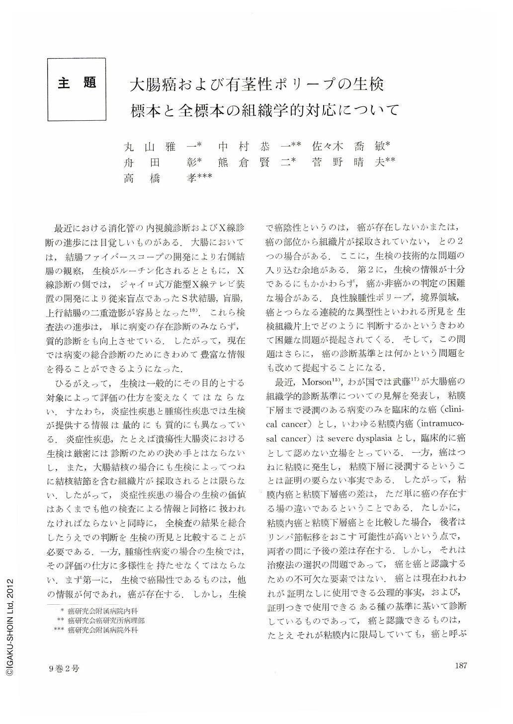
最近における消化管の内視鏡診断およびX線診断の進歩には目覚しいものがある.大腸においては,結腸ファイバースコープの開発により右側結腸の観察,生検がルーチン化されるとともに,X線診断の側では,ジャイロ式万能型X線テレビ装置の開発により従来盲点であったS状結腸,盲腸,上行結腸の二重造影が容易となった10).これら検査法の進歩は,単に病変の存在診断のみならず,質的診断をも向上させている.したがって,現在では病変の総合診断のためにきわめて豊富な情報を得ることができるようになった.
ひるがえって,生検は一般的にその目的とする対象によって評価の仕方を変えなくてはならない.すなわち,炎症性疾患と腫瘍性疾患では生検が提供する情報は量的にも質的にも異なっている.炎症性疾患,たとえば潰瘍性大腸炎における生検は厳密には診断のための決め手とはならないし,また,大腸結核の場合にも生検によってつねに結核結節を含む組織片が採取されるとは限らない.したがって,炎症性疾患の揚合の生検の価値はあくまでも他の検査による情報と同格に扱われなければならないと同時に,全検査の結果を総合したうえでの判断を生検の所見と比較することが必要である.一方,腫瘍性病変の場合の生検では,その評価の仕方に多様性を持たせなくてはならない.まず第一に,生検で癌陽性であるものは,他の情報が何であれ,癌が存在する.しかし,生検で癌陰性というのは,癌が存在しないかまたは,癌の部位から組織片が採取されていない,との2つの場合がある.ここに,生検の技術的な問題の入り込む余地がある.第2に,生検の情報が十分であるにもかかわらず,癌か非癌かの判定の困難な場合がある.良性腺腫性ポリープ,境界領域,癌とつらなる連続的な異型性といわれる所見を生検組織片上でどのように判断するかというきわめて困難な問題が提起されてくる.そして,この問題はさらに,癌の診断基準とは何かという問題をも改めて提起することになる.
A histo-pathological study was made on one-to-one correspondence in histological finding of biopsied, and operated or polypectomied material, based on 151 lesions of carcinoma and 46 lesions of pedunculated polyp of the large intestine which were experienced in the period of 2 years and 9 months from Jan. 1971 to Sep. 1973 in the Cancer Institute Hospital.
Result:
1. Of the 151 lesions of carcinoma, including 10 lesions of sessile early carcinoma (the definition of early gastric cancer was adopted), biopsy was positive in 142 lesions, regardless of their histological type. Those comprise 94.0% of all lesions biopsied and operated.
2. One-to-one correspondence in histological finding of biopsied and operated material of carcinoma was studied. Histological type of biopsied material (adenocarcinoma papillotubulare, adenocarcinoma muconodulare, and normal mucosa) was contrasted with that of operated material. Normal mucosa means that biopsy is negative for carcinoma. Histological finding of the operated material was divided into three types; adenocarcinoma papillotubulare, adenocarcinoma papillotubulare, partly muconodulare, and adenocarcinoma predominantly muconodulare. Of the 137 operated materials diagnosed as adenocarcinoma papillotubulare, the histological type of 128 materials corresponded to that of the biopsied material, and the 9 of them revealed normal mucosa. Of the 9 operated materials diagnosed as adenocarcinoma papillotubulare, partly muconodulare, the diagnosis of the biopsied material was adenocarcinoma papillotubualre in 8 lesions and adenocarcinoma muconodulare in 1 lesion. On the other hand, of 5 operated materials diagnosed as adenocarcinoma predominantly muconodulare, the diagnosis of biopsied material was adenocarcinoma muconodulare in 4 lesions and adenocarcinoma papillotubulare in 1 lesion.
3. A histological basis of atypia (benign, borderline and malignant) was employed and 46 lesions of pedunculated polyp were classified into those three groups. Twenty-four lesions were diagnosed as benign; 10 lesions were diagnosed as borderline and 12 lesions were diagnosed as malignant.
4. Histological finding of biopsied material of 46 pedunculated polyps was contrasted with that of the total material operated or polypectomied. When both biopsied and operated materials consist of more than two different varieties of atypia (benign, borderline, and malignant), their final diagnosis was made by adopting the severest one, regardless of their qualitative difference in each atypia.
The histological finding of the biopsied material completely corresponded to that of the total material diagnosed as benign (24 lesions) and those diagnosed as borderline (10 lesions). However, in 12 total materials diagnosed as malignant, the diagnosis of the biopsied material was benign in 1 lesion and borderline in 3 lesions. The biopsy finding of the remaining 8 lesions correponded to that of the total materials diagnosed as malignant.
In conclusion, the histological finding of the biopsied materials correponded quite well to that of the operated materials in case of evident carcinoma and pedunculated polyp with uniform histology. However, the biopsied material did not always reflect the every aspect of the total materials in case of pedunculated polyp with mixed histology. Therefore, endoscopic polypectomy would be advisable for the strict histological evaluation of a pedunculated polyp.

Copyright © 1974, Igaku-Shoin Ltd. All rights reserved.


