Japanese
English
- 有料閲覧
- Abstract 文献概要
- 1ページ目 Look Inside
- サイト内被引用 Cited by
胃癌のX線・内視鏡診断技術の著しい進歩によって,これまでに数多くの早期胃癌が発見され,手術切除されてきている.それとともに,いろいろな型の早期胃癌例にも遭遇するようになった.その1つに,粘膜面における癌の拡がりが明瞭でないものがある.このような胃癌の手術にあたっては,その噴門側境界がよく問題9)28)になる.いうまでもなく,粘膜内での癌浸潤の境界がはっきりしない病変(以下Ⅱb様病変と表現)に対しては,その噴門側境界を術前に可能な限り明らかにしておくことが必要である.そのためには,X線診断に際して病変の噴門側境界部がどのような所見として描写されているか,また,どのような所見に注意して境界部の診断を行なえばよいか,という問題の解明が重要である.
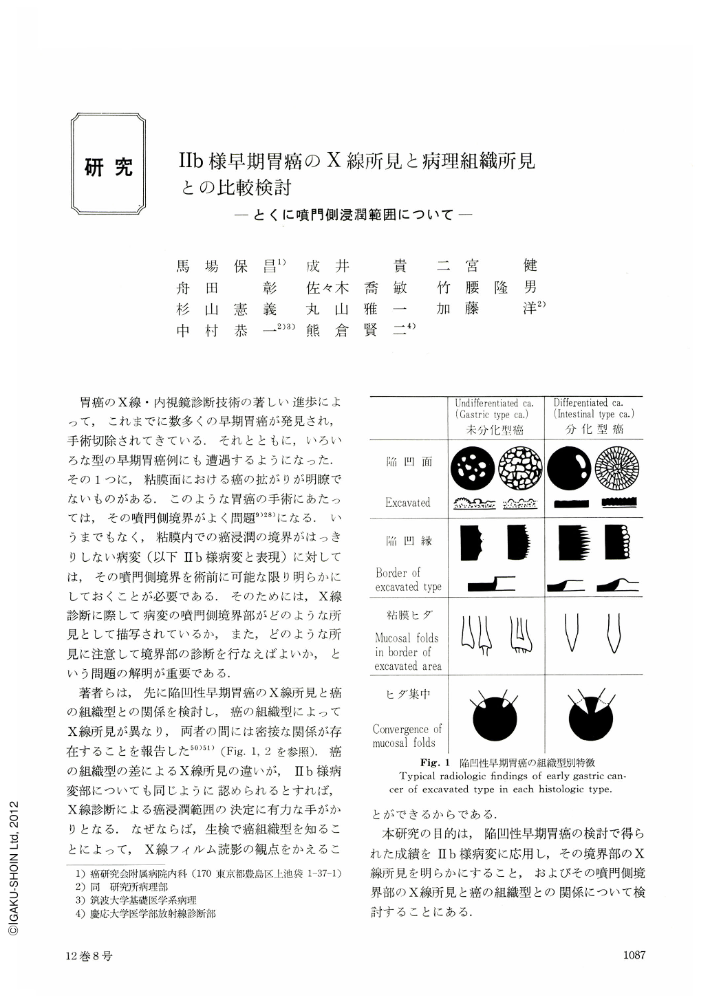
著者らは,先に陥凹性早期胃癌のX線所見と癌の組織型との関係を検討し,癌の組織型によってX線所見が異なり,両者の問には密接な関係が存在することを報告した50)51)(Fig. 1,2を参照).癌の組織型の差によるX線所見の違いが,Ⅱb様病変部についても同じように認められるとすれば,X線診断による癌浸潤範囲の決定に有力な手がかりとなる.なぜならば,生検で癌組織型を知ることによって,X線フィルム読影の観点をかえることができるからである.
The above mentioned study was conducted in order to define radiologically a boundary between Ⅱb-like lesion and the normal surrounding mucosa, based upon 71 such cases which had been obtained in the period of 11 years from 1965 to 1976 at the Cancer Institute Hospital. Especially, an analysis was focused upon correlation between radiologic sign and histologic type of the lesion at its proximal boundary. For analysis of the radiologic findings double contrast radiograph were mainly used. Histologic type of gastric carcinoma was divided into two basic types, namely differentiated carcinoma (Intestinal type) and undifferentiated carcinoma (Gastric type). Following results were obtained.
1. The histologic type was found to be related closely to the radiologic sign in 60% of the Ⅱb-like lesions. In such cases, the proximal boundary was easily defined. 2. A linear shadow of barium which was retained in between the mucosal folds was found to be an aid for defining the proximal boundary. 3. In the lesions which showed characteristic radiologic sign of each histologic type with easily defined boundary, histologic examination revealed numerous minute erosions and these erosions were found more frequently in the undifferentiated carcinomas than the differentiated carcinomas. 4. In addition to defining the proximal boundary for determining surgical cut line, an attention should be paid to a presence of a satellite focus which was located whithin 1 cm of the surgical stump, because 15 such foci were discovered in 125 cases of multiple cancers which were experienced in the recent 6 years.
Copyright © 1977, Igaku-Shoin Ltd. All rights reserved.