Japanese
English
- 有料閲覧
- Abstract 文献概要
- 1ページ目 Look Inside
チリにおける胃癌の頻度は,日本と同様に高いが,進行した胃癌が多く,治療成績も不良であった.近年めざましい進歩をとげた日本の胃診断法をとり入れて,チリーにおいて初めて,Ⅲ+Ⅱc型早期胃癌を発見したので報告する.
PROLOGUE
The incidence of carcinoma of the stomach among the Chilean population is as high as that in the Japanese. In the former, the diagnosis was made usually in an advanced stage of the disease consecuently the therapeutic results have been poor. With the introduction of the gastrodiagnostic method that has shown a brilliant progress in Japan in recent years, a type Ⅲ+Ⅱc carcinoma of the stomach has been discovered as the first case in Chile. This communication describes the case.
Case A72-year woman. She complain of epigastralgia for the past three years, occurring seasonally, and was admitted to the Hospital Salvador because of pain and significant loss of weight. Gastroscopy (Dr. Jaime Klinger) Jan. 1970.
In the angular region of the stomach a shallow ulceration in an elevated zone was observed, the mucosa in the surrounder area was distorted. The folds were interrupted.
Conclusion: Gastric Cancer.
X-Ray Finding (Dr. Ronban):
X-ray examination of the stomach (Dr. Ronban)
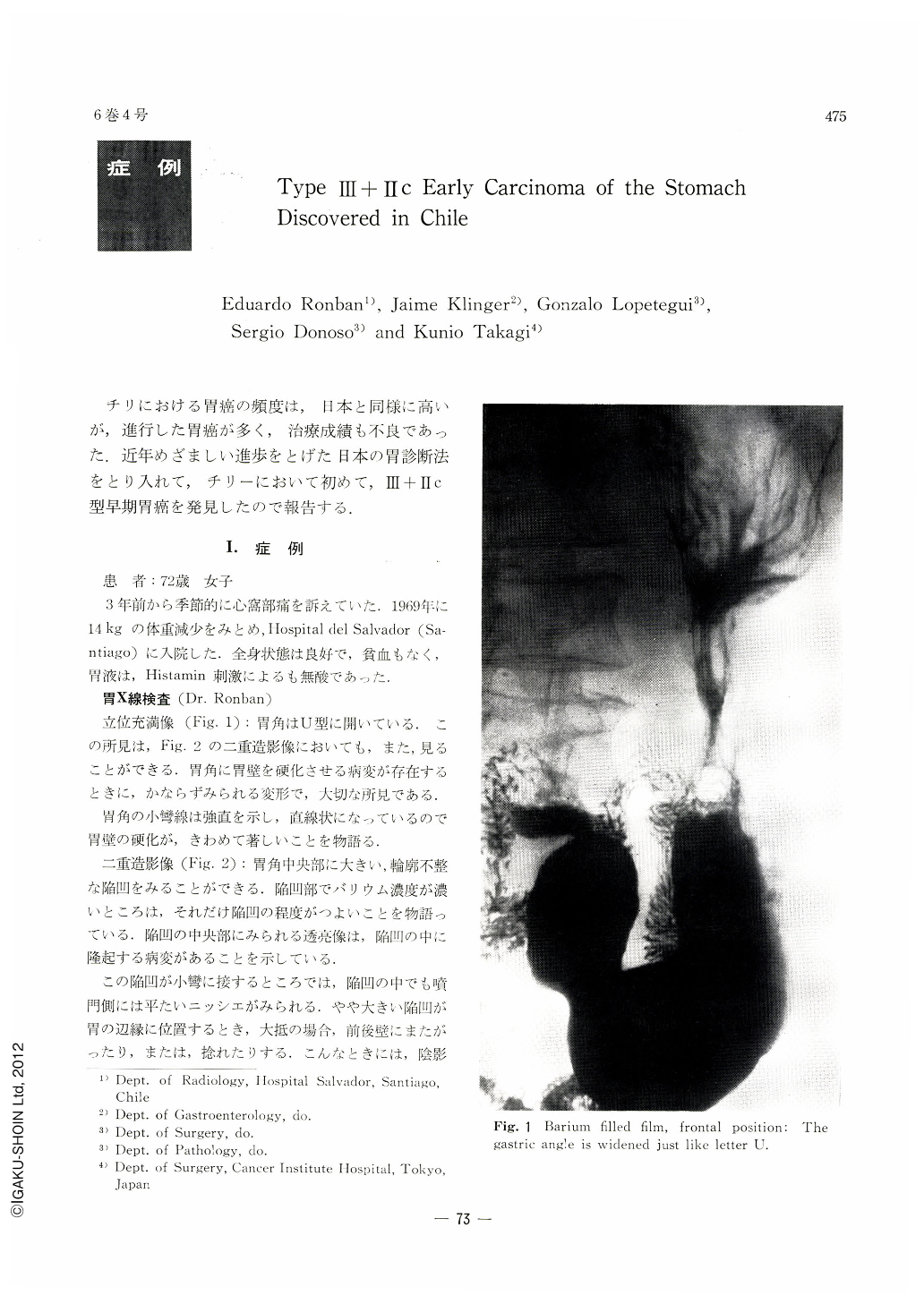
Upright, barium-fiilled picture (Fig. 2): The gastric angle is widened like the letter U. This finding is likewise seen in the double contrast picture in Fig. 2. It is a very important finding seen whenever a lesion making the gastric wall rigid exists at or close to the gastric angle. The contour of the lesser curvature around the angle is stiff and presents straight line, indicating that rigidity of the wall there is remarkable.
Double contrast picture (Fig. 2): At the incisura can be seen a large depression of irregular outline. Denser barium shadow within it shows that the degree of depression is so much deeper. A radiolucency in its center bespeaks a protruding lesion within it.
At a place where a part of the depression located on the posterior wall comes into contact with the lesser curvature, a flat niche is observed in the oral part of the depression. A large depressed lesion, located at or near the gastric border, is usually streched to both sides of the wall, sometimes in a twisted fashion. A shadow defect is to be observed then. In the present instance the lesion is not twisted, seen clearly along the gastric contour as a complete profile view. It accounts for the formation of a niche clearly visualized in this picture.
Abnormal mucosal tips are also well seen within the depression on the posterior wall around the gastric border. In the pyloric portion of the depression the mucosal tips are slightly tortuous and rigid. The outline of the folds is irregular on both sides. Some of them have ceased abruptly. Those in the cardiac portion of the depression are swollen, and some are confluent. Its cardiac margin looks as if a little protruded.
The contour of a depressed lesion is made all the more conspicuous accoding as its marginal protrusion becomes distinct. Such a picture as this dose indicate a higher degree of depression among lesions of superficially depressed type early gastric cancer.
Finding of Va-type gastrocamera (Dr. Lopetegui)
A slender, irregular, shallow ulcer was found on the gastric wall in an area extending from the lesser curvature at the angle over posterior wall Fig. 3. The mucosa at the ulcer margin on the posterior wall side was depressed circularly, surrounding the ulcer.
Subtotal gastrectomy with omentectomy was performed with the diagnosis of gastric carcinoma, on March 4, 1970. (Operator: Dr. Lopetegui) Gross Findings (Fig. 4): A slender, zonal depression of mucosa at the ulcer margin on the posterior wall side, surrounding the ulcer, was in evidence and the mucosa within the sphere of 1.5cm diameter on the anterior wall side was slightly disclored and its surface finely granular. Microscopic Findings: Three representative sections of the stomach from the resected specimen were obtained (Fig. 5, 8 a b). There was no carcinomatous growth on the ulcer bottom, reaching the proper muscle layer, but there was evidence of infiltration of adenocarcinoma mucocellulare et tubulare into the mucosa at the ulcer margin (Fig. 6, 7). The adenocarcinomatous growth was found infiltrating the submucosa, (tissue section #2.) Dr. Takagi who happened to be in Chile prepared step-sections of the tissue from the resected specimen looking for the extention of the cancer (Fig. 9). The carcinoma within the mucosa was found to exist primarily on the anterior wall side, surrounding the ulcer, and submucosal infiltration was evident at the ulcer margin. There was no evidence of carcinomatous growth on the ulcer bottom, however. Metastatic carcinomas were found in two lymph nodes at the lesser curvature.
SUMMARY & CONCLUSION
A statistic survey on deaths due to carcinomas macle by Segi has evidenced the incidence of gastric carcinoma to be the highest among the Chilean and the Japanese populations in the world. The recent, rapid progress in the various diagnostics of the stomach in Japan has contributed to a considerable extent to the discovery of gastric carcinomas, especially those in the early stage, and, in consequence, cases discovered as early carcinomas of the stomach account for as many as about 20 per cent of all cases operated on for gastric carcinoma in Japan. Results of surgical treatmant for early carcinoma of the stomach are also remarkably satisfactory; as many as 90 per cent of patients treated for early carcinoma usually survive for not less than five years. The advanced in diagnostics has been highlighted all over the world and the Chilian physicians with this diagnostic technique have succeeded in the detection of Ⅲ+Ⅱc type early carcinoma of the stomach. The case herein described lacks suffcient data and the whitish tone of colors encountered with the photogastrograms was due to the use of a stale role film. The roentgenograms, gastrocameras and findings of the resected gastric specimen have led to the diagnosis of gastric carcinoma but the ings on microscopic examination of the initial three cut sections from the resected specimen suggested a benign lesion, thereby making the physicians engaged in the clinical examinations perplexed.
Drs. Donoso and Takagi re-examined the tissue slides microscopically and made diagnosis of early gastric carcinoma with submucosal infiltration; thus the initial clinical diagnosis was finally accepted. Such an event as this had been experienced from time to time in Japan a decade ago when early carcinomas of the stomach had barely begun being discovered, and the disparity in diagnostic views between the clinician and the pathologist became eliminated by encouraging pathologists to familiarize themselves with the microscopic features of early carcinomas of the stomach.
Medical interchange between Chile and Japan has just begun with respect to gastro-diagnostic techniques and We wish the medical interchange between the two countries to progress in all the more profound harmony.
The authors with to thank Prof. Hikoo Shirakabe of Juntendo University for his invaluable instruction of x-ray findings.
Copyright © 1971, Igaku-Shoin Ltd. All rights reserved.