Japanese
English
- 有料閲覧
- Abstract 文献概要
- 1ページ目 Look Inside
1.症例
患者:62歳 男
主訴:特に述べることはない.
現病歴:母が胃癌死亡のため,7年前胃精検を受けたが,当時は異常なしと言われた.約3カ月前頃から食欲はあるが,食後時々胃部不快感があり,時に軽い疼痛があったので,昭和42年12月6日本院に受診し,加療により症状は間もなく消褪したが,X線,内視鏡検査の結果胃前庭部に隆起性病変を指摘され,昭和43年1月8日手術のため本院に入院した.
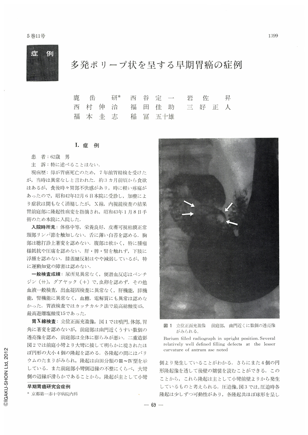
As a result of various examinations, a protruding lesion was found in the gastric antrum of a man who visited the out-patient clinic for a thorough check-up of his stomach. Under the diagnosis Of gastric cancer gastrectomy was performed. The lesion was an early gastric cancer having multiple polypoid appearance. General examinations including function tests of the liver, spleen and kidney were normal. His stool was positive for benzidine test (++) as wll as for guajac test (+). Examination of the gastric contents by Katsch Kalk's method revealed total acid 45° and free hydrochloride acid 15°. At x-ray examination, several translucencies were recognized in the antrum in an upright, bariumfilled picture. These were further confirmed as four protrusions of varying size both by double contrast and compression studies. Gastrocamera revealed four pedunclated elevations in the antrum around the anterior wall near the lesser curvature. The color and luster of the surface of the protursions showed hardly any difference from those of the normal mucosa. However, biopsy disclosed the existence of adenoearcinoma in these elevations. Structural at- ypia was not so apparent, but cellular atypia was seen in a marked degree. These protrusions measured, from oral to diatal, 20×20, 30×20, 10×20 and 10×10 mm, respectively. They were all polypoid protrusions having short stalk. Histopathologically, all four elevations belonged to adenocarcinoma tubulare showing marked papillary hyperplasia on the surface. No malignant finding was seen in the border areas of these elevations. Cancer infiltration remained all within the mucosa. They were of type I early cancer.

Copyright © 1970, Igaku-Shoin Ltd. All rights reserved.


