Japanese
English
- 有料閲覧
- Abstract 文献概要
- 1ページ目 Look Inside
初回のレ線診断で良性潰瘍と診断したが,内視鏡検査では広範な広がりのⅡc病変内に,BorrmannⅡ型を合併する進行癌と診断し,術後これを確認した症例を報告する.本例は胃疾患診断における綜合診断の必要性と,多方向よりのレ線撮影の有用性を示すものである.
The patient: a 53-year-old male
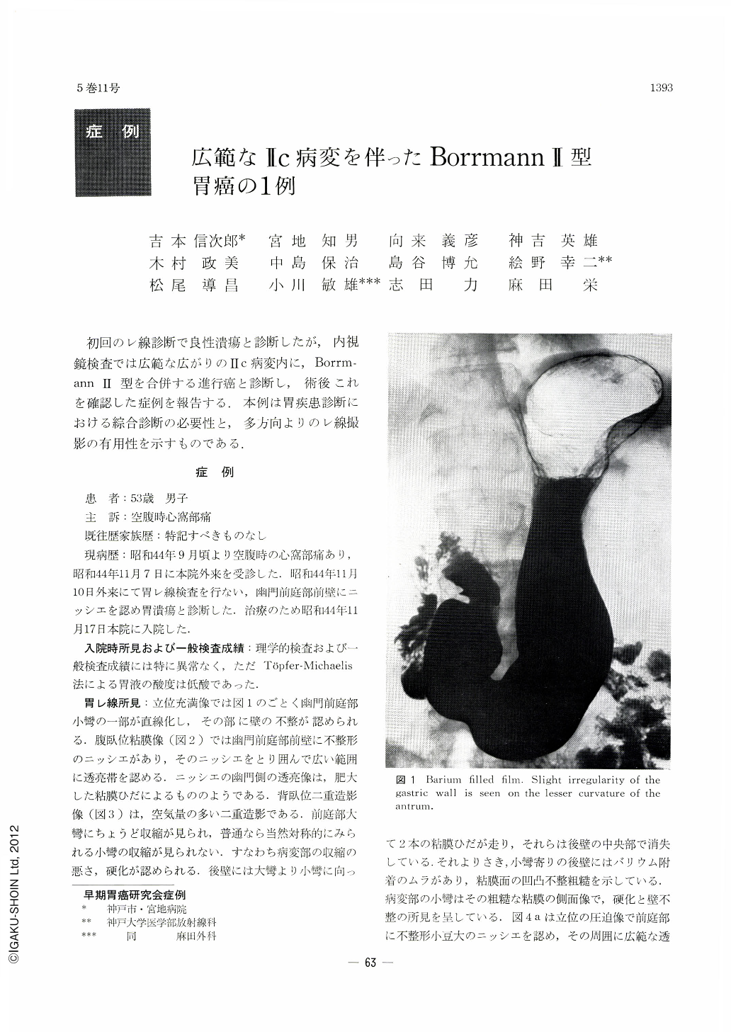
He visited the authors’ hospital complaining of epigastric hunger pain of three months’ duration. At x-ray examination, an irregular small niche the size of a red bean was seen on the anterior wall of the pyloric antrum. Extensive translucent zone was seen around it. Its outer margin was sharply defined, and in the pyloric side it was visualized as of flower-petal-like appearance. In the prone double contrast study in the second oblique projection, the niche was seen as an excavated niche. Gastroscopically, an ulcer of irregular shape was observed on the anterior wall of the pyloric antrum in addition to an extensive, abnormally engorged area surrounding the ulcer. Two swollen mucosal folds were seen to converge toward it from the pyloric side. Operation was performed under the diagnosis of aclvanced carcinoma of the stomach. The resected stomach showed on the anterior wall of the pyloric antrum an ulcer, measuring 1.5×1.5 cm, shaped like a figure of snowman. It was almost round with a partial protrusion on the greater side, having a notable embankment around it. The mucosa around the ulcer, aboutcm in extent, showed shallow, erosive depression, indicative of Ⅱc lesion. Pathologically, the ulcer belonged to Borrmann II type advanced carcinoma invading the muscular coat. The depressed area around it was IIc, mostly localized within the mucosal layer with partial encroachment into the submucosa. Its histological diagnosis was adenocarcinoma (CAT II, SAT 3, INFβ) tubulare, pm ly1, Vo, So, ow (-), aw (-)

Copyright © 1970, Igaku-Shoin Ltd. All rights reserved.


