Japanese
English
- 有料閲覧
- Abstract 文献概要
- 1ページ目 Look Inside
胃癌の発生母地として,潰瘍,ポリープ,胃炎のほか,迷入組織もその一つに古くから考えられている.迷入組織としては主に腸上皮,食道粘膜上皮および膵組織がとりあげられているが,迷入膵組織から癌化したと思われる症例は,ごく早期のものであっても癌病巣は胃壁深層にみられることが多い.
最近筆者らは迷入膵が母地となったと思われ,しかも粘膜層のみに限局したⅡc型早期胃癌の極めて珍らしい症例を経験したので報告する.
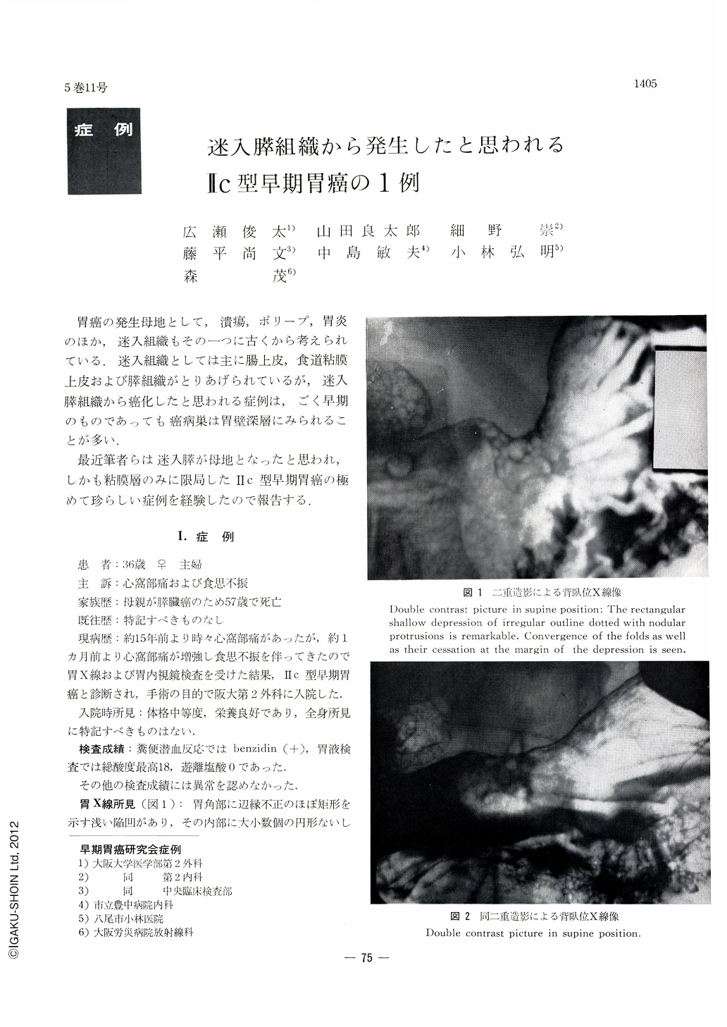
The patient is a 36-year-old housewife complaining of epigastralgia and loss of appetite. Her mother died of cancer of the pancreas. At x-ray examination, an almost rectangular shallow depression of irregular outline was seen at the gastric angle. Several small round protrusions were seen within the depression together with convergence and abrupt cessation of the mucosal folds around it. It was presumed then to be Ⅱc (+Ⅱa) type early gastric cancer with possible submucosal invasion. Endoscopical pictures revealed an irregular ulcer at the gastric angle a little on the side of the posterior wall. It was covered with whitish exudate. A clearly circumscribed shallow depression was seen on the oral side of the ulcer. The depressed area had nodular protrusions of varying size within it. On its anal side was seen edematous mucosal embankment of smooth surface. Endoscopically, the lesion was thus suspected to be Ⅱc+Ⅲ type early cancer with high possibility of submucosal infiltration. The resected stomach disclosed on the posterior wall at the level of the gastric angle a little on the anal side a shallow depression of irregular contour having at places deeper cavities and islet-like protrusions. Slight mucosal convergence was also recognized. The lesion was next considered to be IIc+III type early cancer with possible submucosal invasion. Histologically, the shallow depression belonged to early cancer with its infiltration limited within the mucosa. Most of it was signet ring cell carcinoma, gradually replaced by tubular adenocarcinoma in the deeper layers. The deep cavities mostly belonged to U1-I with partial Ul-II type ulcer scar. Beneath the IIc lesion, mostly located in the muscularis propria, were tissues of aberrant pancreas identical in structure with proper tissues of the pancreas. A fraction of them reached through the lamina propria up to the submucosa. Cell atypia was manifest in a part. The cancer lesion in the deeper region of the mucosa showed cells with eosinophile granules and cells having the same structure of centroacinous cells seen in the normal tissues of the pancreas. The afore-mentioned part of the aberrant pancreas, reaching up to the mucosal surface, were located almost in the center of the IIc lesion.
These findings naturally led the authors to the conception of Ⅱc type early gastric cancer arising from the aberrant pancreas. Such a case, unlike those cases originating from the aberrart pancras as described in the literature, is considered to be of a very rare occurrence.

Copyright © 1970, Igaku-Shoin Ltd. All rights reserved.


