Japanese
English
- 有料閲覧
- Abstract 文献概要
- 1ページ目 Look Inside
- サイト内被引用 Cited by
はじめに
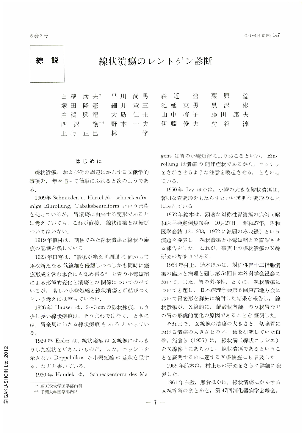
線状潰瘍,およびその周辺にかんする文献学的事項を,年々追って簡単にふれると次のようである.
1909年Schmieden u. Härtelが,schneckenförmige Einrollung,Tabaksbeutelformという言葉を使っているが,胃潰瘍に由来する変形であるとは考えていても,これが直接,線状潰瘍とは結びついてはいない.
1919年植村は,剖検でみた線状潰瘍と線状の瘢痕の記載を残している.
1923年田宮は,“潰瘍が絶えず周囲に向かって逐次新たなる筋線維を侵襲しつつしかも同時に瘢痕形成を営む場合にも認め得る”と胃の小彎短縮による形態的変化と潰瘍との関係についてのべているが,著しい小彎短縮と線状潰瘍とが結びつくという考えには至っていない.
1926年Hauserは,2~3cmの線状瘢痕,もう少し長い線状瘢痕は,そうまれではなく,ときには,胃全周にわたる線状瘢痕もあるといっている.
1929年Eislerは,線状瘢痕はX線像にはっきりした症状をださないものだ,また,ニッシエを示さないDoppelulkusが小彎短縮の症状を呈する,などと書いている.
1930年Haudekは,Schneckenform des Magensは胃の小彎短縮によりおこるといい,Einrollungは潰瘍の随伴症状であるから,ニッシェをさがさせるような注意を喚起させる,ともいっている.
1950年Ivyほかは,小彎の大きな鞍状潰瘍は,著明な胃変形をもたらすといい著明な変形のことにふれている.
1952年鈴木は,顕著な対称性胃潰瘍の症例(昭和医学会定例集談会,10月27日,昭和27年,昭和医学会誌12:203,1952に演題のみ収録)という演題を発表し,線状潰瘍と小彎短縮とを直結させる報告をした.これが,事実上の線状潰瘍のX線研究の始まりである.
1954年村上,鈴木ほかは,対称性胃十二指腸潰瘍の臨床と病理と題し第54回日本外科学会総会において,また,胃の対称性,とくに,線状潰瘍にっいてと題し,日本病理学会第6回東部地方会において胃変形を詳細に検討した結果を報告し,線状潰瘍が,X線的に,蝸殻状内醗,のう状胃などの胃の形態的変化の原因であることを証明した.それまで,X線像の潰瘍の大きさと,切除胃における潰瘍の大きさとの不一致を研究していた白壁,熊倉ら(1955)は,線状溝(線状ニッシエ)をX線像上にあらわし,線状潰瘍であるということを証明するのに適するX線検査にも言及した.1959年鈴木は,村上らの研究をさらに詳細に発表した.
1961年白壁,熊倉ほかは,線状潰瘍にかんするX線診断のまとめを,第47回消化器病学会総会,アジヤ消化器病学会に報告した.
1962年増田ほかは,ミュンヘンで行なわれた第2回世界消化器病学会においてClinical,fluroscopic and endoscopic observations on the symmetric ulcers(Linear and Kissing ulcers)of the stomachと題し,さらに進歩した内容の診断学を発表し欧米の学者を啓蒙し,わが国の消化器病学者に自信を与えた.
1963年BochusのGastroenterologyに村上,白壁の論文が引用された.
1965年Frick, W.が分担執筆したLehrbuchder Roentgendiagnostikに白壁の論文が引用され,彼自身の考案による胃変形図がでた,しかし,1961年にFrikは,すでに熊倉の作った線状潰瘍や多発潰瘍の変形図を見て理解しているので,その考え方を多分に取り入れたことは充分に察知できる.
胃内視鏡的には,1937年Gutzeitが,すでにみている.胃角の大きな長い潰瘍の経過を観察しているうちに,strichförmige Narbeといい,彼の著書に線状潰瘍の写真をみることはできる.しかし,村上,鈴木の発表以前には,対称性潰瘍,線状潰瘍をとり上げて,これを詳細検討した研究はない.
1958年中島,西沢ほかは,線状潰瘍の胃カメラ診断について発表した.
その後,線状潰瘍にかんする多数の貴重な発表をみたわけであるが,われわれが敬意を払わなくてはならない研究も多い.例えば,川井(京府大増田内科)による潰瘍の経過観察からみた線状潰瘍,増田(東北大山形内科)のX線学的統計観察,岡部(九大勝木内科)のX線診断と内視鏡診断との詳細な対比研究などである.
線状潰瘍の定義にも問題がある.
村上は,はじめから3cm以上のものを線状潰瘍としている.これは,暗に病因論を考慮してのことであったであろうが,今にして思えば,慎重な取り扱いであったと筆者らは思っている.はじめから線状のもの,潰瘍の経過中に線状の型を呈したもの,また,X線診断の側からいえば,著明な胃変形とそうでないもの,こんな話題の中で占める3cmの意義は,まことに興味があるといえる.
村上,鈴木は,線状潰瘍に円形,または,接吻潰瘍を含むものも線状潰瘍としての分類に含ませている.佐野は,線状潰瘍上に,円形,または接吻潰瘍を含むものは線状潰瘍から除外している.
筆者らのX線診断の立場は,病因論や病理組織学的議論には目もくれずに,ただ一筋に,肉眼的に線状にみえるもの,溝状のもの,X線像上ニッシエとして写るものは,すべて線状潰瘍として取り扱った.なるほど,研究してきた経過中でも,また,いまでも,長い線状潰瘍と,短いものに対する筆者らの気持は,それぞれに違ったものをもっている.しかし,X線診断の側からいえば,どんなに短かくとも線状であることを写しだすのがX線診断であろうと思っている.
The paper deals with 81 cases of linear ulcer, subjected to close examinations both pre and post-operativeiy, out of 88 such cases which underwent x-ray and endoscopic studies at the First Department of Internal Medicine and were operated on the Surgical Department, Chiba Univ., 1955 through 1966. The author discusses the results of the examinations and draws the following conclusions:
1. Histological findings
The incidence of linear ulcer is 17.7%, and 4.9% of it are linear scars. There have been no early gastric cancer existing in the surrounding areas of linear ulcers.
2. X-ray diagnosis
Linear ulcer conspicuously shortens the lesser curvature. Generally speaking, the longer the linear ulcer is, the shorter becomes the distance between the pylorus and the ulcer, and the more marked is the deformity of the stomach. In this paper, the shortening of the lesser curvature is classified into five degrees, ++++, +++, ++, + and -, according to the markedness of the gastric deformity. The symbol, ++++ represents the stomach showing pouch-like deformity and ++, the rightanglecl stomach. Most of cases which fall under the ++++ classification have linear ulcers longer than 75mm. About 50% of them in cases of +++ deformity are between 60 and 70 mm long; about 65 % in ++ deformity are from 30 to 45 mm long, and 83% of + and - are less than 30 mm in length. However, linear ulcers of the gastric body and those chiefly situatecl on the anteriar wall or located parallel to the lesser curvattre anrl exceptions, for the shortening of the lesser curvature is slight in these cases as compared with the length of the linear ulcer. The relation between its length and the shortening of the lesser curvature, as mentioned before, is applicable only to 96.3% of linear ulcer. The discovery rate of niche in it is 76.5%. That of linear niche is 63.0%. The diagnostic accuracy is 69.1%. The deformity of the stomach due to the shortening of the lesser curvature is an important finding for the x-ray diagnosis of linear ulcer. Double contrast method in supine position is most effective in demonstrating a linear niche.
3. Combination of x-ray and endoscopic examinations.
By the combined use of x-ray and endoscopic examinations, diagnostic accuracy has increased up to 93.5%. In cases in which marked deformity of the stomach is recognized or a long linear ulcer is demonstrated, the diagnostic accuracy of x-ray examination is superior to endoscopic study, while the latter surpasses the former in cases in which there is only a slight deformity or a short linear ulcer.
4. Diagnostic limitations
A lesion 1.5mm deep and about 10 mm long can be diagnosed by x-ray. A lesion less than 0.5mm in depth is difficult to interprete. Endoscopically, it is difficult to obtain direct findings of the stomach having marked deformity.

Copyright © 1970, Igaku-Shoin Ltd. All rights reserved.


