Japanese
English
- 有料閲覧
- Abstract 文献概要
- 1ページ目 Look Inside
はじめに
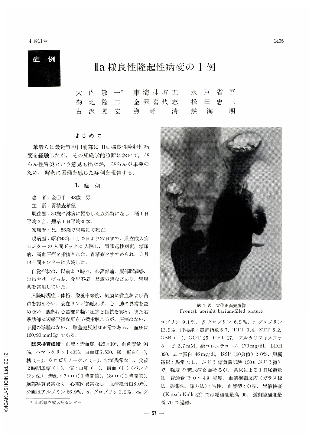
筆者らは最近胃幽門前部にⅡa様良性隆起性病変を経験したが,その組織学的診断において,びらん性胃炎という意見も出たが,びらんが単発のため,解釈に困難を感じた症例を報告する.
A man 48 years of age was found to harbor a protruded lesion in the pyloric antrum of his stomach. He eventually underwent gastrectomy because the result of a thorough examination was such that it was impossible to exclude malignancy although the lesion was considered in all probability of benign nature.
Resected stomach revealed that on the lesser curvature of the pyloric antrum there was an irregular-shaped protrusion, measuring 17 by 16mm, with its surface rough and uneven. In its center a little oralwarcl there was an engorged depression as of an erosion, but the protrusion itself was not so hard to the touch.
Histologically, the lesion was caused by hyperplasia of the mucosa as well as hypertrophy of the walls of the blood vessels in the submucosa, muscle layer and the subserosa. The mechanism of its development remained unknown. Further detailed study of the lesion seems to indicate that it might be hypertrophy of the gastric wall of inflammatory nature, but it is also impossible to deny that the lesion has developed as an inflammatory reaction of the gastric wall against tumorous proliferation, or changes of the gastric wall due to hypertrophy of blood vessels. A sequence of cause and effect remains unestablished in this case.
If there had been multiple protrusions with uneven depressions, the diagnosis of gastritis erosiva could easily have been reached at. A solitary protrusion of benign nature is hard to interprete.
In the clinical diagnosis of a protruded type of early gastric cancer, especially of Ⅱa type, its most characteristic feature is said to consist in sharp uprising of the margins of the lesion with its surface formed like a plateau; however, it is very noteworthy that there do exist, though very few in number, benign protrusions with small erosions as in this case. Though less diversified in their color and of less hardness as compared with malignant lesions, their surface is more or less uneven, hardly to be discriminated from malignancy by gross observation alone. Biopsy and cytologic diagnosis are essential in such a case. Malignancy was at first suspected endoscopically in the case here presented, biopsy was difiicult to perform due to the site of the lesion. Cytologic examination was negative for cancer.

Copyright © 1969, Igaku-Shoin Ltd. All rights reserved.


