Japanese
English
- 有料閲覧
- Abstract 文献概要
- 1ページ目 Look Inside
患 者:伊○忠○ 63歳 男
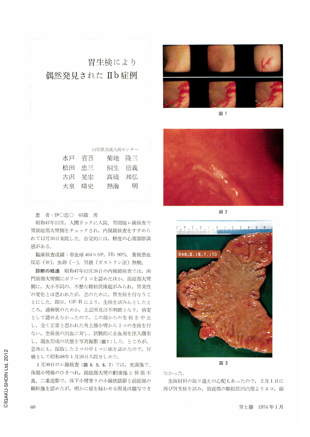
昭和47年12月,人間ドックに入院.胃間接レ線検査で胃前庭部大彎側をチェックされ,内視鏡検査をすすめられて12月26日来院した.自覚的には,軽度の心窩部膨満感がある.
At an indirect radiography of the stomach taken in the course of periodic check-up of his health, a 63-year-old man was noticed to have atrophic, hyperplastic gastritis of the antrum. He was then referred to us for thorough examination. Quite accidentally biopsy specimens were taken from an area on the lesser curvature above the angle, where Ⅱb subtype early cancer was detected. The lesion was adenocarcinoma mucocellulare, measuring 60×55 mm and located on the anterior wall of the middle part of the stomach. Its depth was m. The mucosa in this area was flat and the areae gastricae were indistinct. The non-cancerous part on the posterior wall was also symmetrically ironed out, so that discrimination from a picture of atrophic gastritis was difficult. Discoloration of the mucosa was seen over an area corresponding almost to that of cancer. In barium-filled picture was seen a stricture on the lesser curvature of the lower body, but as we were unable to take a good double contrast picture of the anterior wall, any finding suggestive of cancer was unobtainable. Even retrospective study of endoscopic pictures taken in close-up view did not yield much for us to grasp the whole view of the cancer lesion, although it was not impossible to notice slight discoloration of the mucosal surface. At all events, there is nothing for it at present for the detection of such a case as this but luck. At least, however, we should work hard to detect even Ⅱb, however difficult it may be.

Copyright © 1974, Igaku-Shoin Ltd. All rights reserved.


