Japanese
English
- 有料閲覧
PPV購入案内
1,320円 (1,200円+税10%) こちらは、1507ページから1509ページの文献です。購入には医書.jp本会員登録が必要です。
会員登録完了後に改めて上記「購入サイトへ」を選択してください。
または「購入サイトへ」選択後に「購入ログイン」、「新規会員登録」の順に進んでください。
会員登録について詳しくはをご確認ください。
- Abstract 文献概要
- 1ページ目 Look Inside
患 者:S. H. 65歳 女
病 歴:3年来糖尿病で通院し,Controlは良好であった.約20日前右上腹部に疝痛発作があり,2日前より悪感,発熱と共に黄疸が出現し入院.その後症状は比較的急速に消失し,1年後の現在も経過良行である.
In the course of choledochitis a nodular tumor of inflammatory origin in the papilla of Vater was seen in a 65-year-old woman. For three years she had ambulatory treatment for diabetes mellitus, which was under good control. About 20 days before she had a bout of colic in the right upper quadrant. She was then hospitalized because icterus appeared beside chill and fever. After admission the symptoms went away fairly repidly. As of one year after operation she has a favorable course.
Results of examinations showed that directly after admission the liver was palpable one finger and a half's breadth below the costal arch. The right hypochondrium was tender. Blood showed increase of white blood cells. Serum Wassermann's test was positive. Serum bilirubin was 6.3 mg/dl (direct 4.2; indirect: 2.1). SGOT was 54 units with SGPT 75 units. Alkaline phosphatase was 55 KA units, LDH being 330 units, TTT 4 units, Co. R(1). Serum cholesterol was 242 mg/dl, with CRP positive. Blood glucose level was 110 mg/dl.
X-ray films of the chest showed an inactive shadow in the lower field of the right lung.
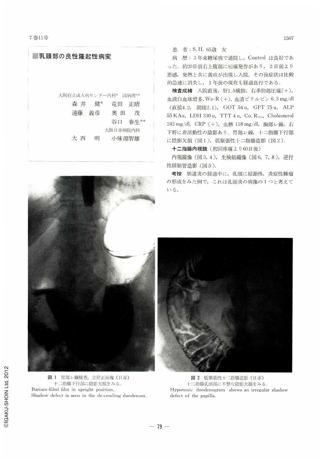
Roentgenogram of the upper digestive tract disclosed a shadow defect in the second portion of duodenum (Figure 1). Hypotonic duodenography is shown in Figure 2.
Duodenofiberscopic examination (60 days after the initial pain). Endoscopic pictures (Figures 3 and 4). Histology of biopsied specimen (Figures 6, 7 and 8). Retrograde pancreatocholedochography (Figure 5).
Copyright © 1972, Igaku-Shoin Ltd. All rights reserved.