Japanese
English
- 有料閲覧
- Abstract 文献概要
- 1ページ目 Look Inside
- サイト内被引用 Cited by
Ⅰ.十二指腸のX線検査手技
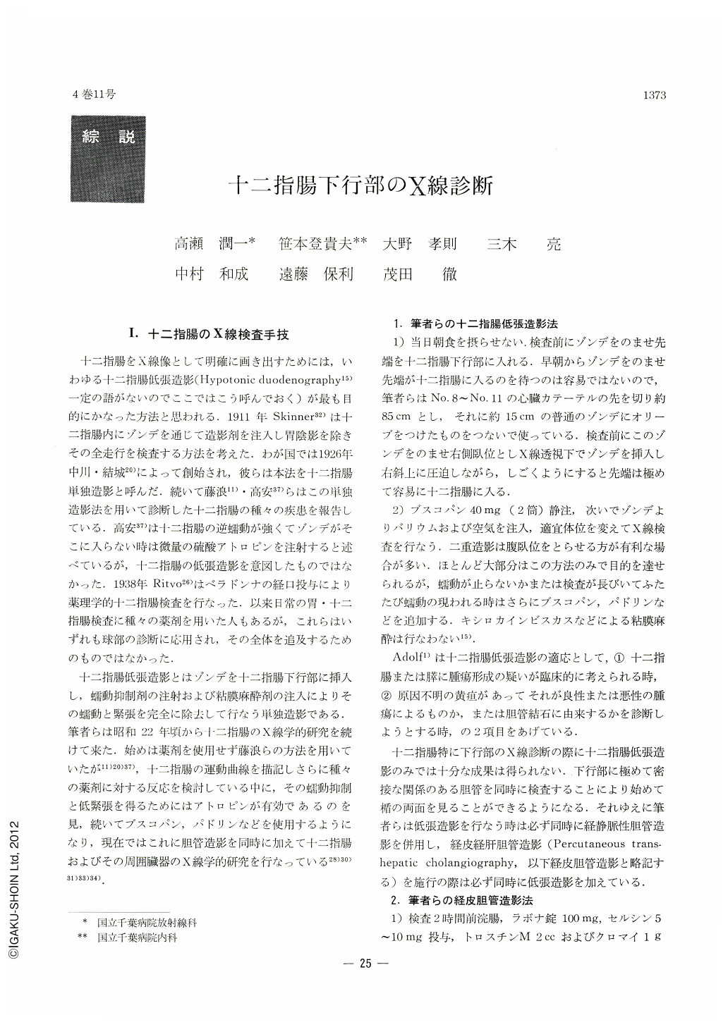
十二指腸をX線像として明確に画き出すためには,いわゆる十二指腸低張造影(Hypotonic duodenography15)一定の語がないのでここではこう呼んでおく)が最も目的にかなった方法と思われる.1911年Skinner32)は十二指腸内にゾンデを通じて造影剤を注入し胃陰影を除きその全走行を検査する方法を考えた.わが国では1926年中川・結城20)によって創始され,彼らは本法を十二指腸単独造影と呼んだ.続いて藤浪11)・高安37)らはこの単独造影法を用いて診断した十二指腸の種々の疾患を報告している.高安37)は十二指腸の逆蠕動が強くてゾンデがそこに入らない時は微量の硫酸アトロピンを注射すると述べているが,十二指腸の低張造影を意図したものではなかった.1938年Ritvo26)はベラドンナの経口投与により薬理学的十二指腸検査を行なった.以来日常の胃・十二指腸検査に種々の薬剤を用いた人もあるが,これらはいずれも球部の診断に応用され,その全体を追及するためのものではなかった.
十二指腸低張造影とはゾンデを十二指腸下行部に挿入し,蠕動抑制剤の注射および粘膜麻酔剤の注入によりその蠕動と緊張を完全に除去して行なう単独造影である.筆者らは昭和22年頃から十二指腸のX線学的研究を続けて来た.始めは薬剤を使用せず藤浪らの方法を用いていたが11)20)37),十二指腸の運動曲線を描記しさらに種々の薬剤に対する反応を検討している中に,その蠕動抑制と低緊張を得るためにはアトロピンが有効であるのを見,続いてブスコパン,パドリンなどを使用するようになり,現在ではこれに胆管造影を同時に加えて十二指腸およびその周囲臓器のX線学的研究を行なっている28)30)31)33)34).
By combining hypotonic duodenography with percutaneous transhepatic cholangiography, x-ray diagnosis of diseases of the duodenum and its adjacent organs can now be made exactly. In this paper are described the technics of both procedures together with such diseases as are within reach of diagnosis by these maneuvers.
The mucosal relief of the duodenum obtained by hypotonic duodenography is a little coarse but usually not to such an extent as to be beyond its conception; the papilla of Vater can be recognized normally in about 60 per cent as and almost circular, protruded silling defect. It swells up according as it is affected by inflammation in the regions of the bile duct and the pancreas. When the muscle of Oddi is ruined by long-standing inflammation, contents of the duodenal lumen may regurgitate up into the bile duct.
Discrimination between benign and malignant tumors of the duodenum is roentgenologically not difiicult, and percutaneous transhepatic cholangiography is of great diagnostic significance because the papilla of Vater is the predilection site of malignant tumors.
Lesions of the pancreas affect direct or indirect changes on the duodenum. Pancreatitis presents itself as dilatation or deformation of the duodenal loop. It may also cause Winding or narrowing of the terminal part of the choledochus followed by dilatation of its upper part. For early detection of pancreatic cancer, deformity of the duodenal loop is less important than findings in the terminal part of the common bile duct visalized by percutaneous transhepatic cholangiography.
Duodenal diverticula, because of the location of their predilection sites, greatly affect the bile duct as well as the pancreas. Of greater impoptance are secondary changes caused by the presence of duodenal diverticula rather than their clinical significance itself. It also has been presented in this paper that lesions in the bile duct and the pancreas can be diagnosed by observation of the duodenal diverticula themselves.

Copyright © 1969, Igaku-Shoin Ltd. All rights reserved.


