Japanese
English
- 有料閲覧
- Abstract 文献概要
- 1ページ目 Look Inside
はじめに
胃のX線診断の進歩は,X線写真と切除標本との比較がその基盤となっている.すなわち,両者の所見を対比してみて,自らの診断技術の拙さを知り,病変の描出や読影能力の向上に努める.この繰返しで,病変の描出や,読影力は進歩し,対比症例のつみ重ねから,X線所見の整理・分類が行なわれ,所見の客観性が確立される.
胃のX線診断を志す人にとって,胃の切除標本は,最上の師であり,鞭である.
一方,十二指腸潰瘍は,胃疾患に劣らぬほど数多い疾患であるのに,そのX線診断は,遅々として進歩しない.原因は,種々であろうが,最大の理由は,師であり鞭である切除標本が,極めて得難いことにあろう.
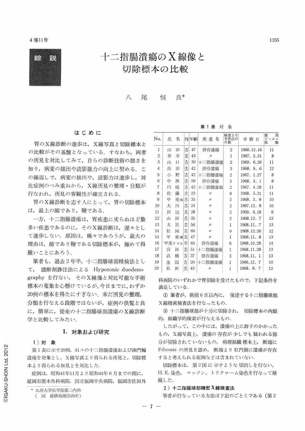
筆者も,過去2年半,十二指腸球部精検法として,遮断剤静注法によるHypotonic duodenographyを行ない,そのX線像と対比可能な手術標本の蒐集を心懸けているが,今日までに,わずか20例の標本を得たにすぎない.未だ所見の整理,分類を行なえる段階ではないが,症例の供覧と共に,簡単に,従来の十二指腸球部潰瘍のX線診断学と比較してみたい.
In order to find diagnostic limit of ulcer of the duodenal bulb, 41 lesions (including ulcer scars) in 20 ulcer of cases that region, surgically resectetl amply enough so that their nature might be examined in detail both macroscopically and microscopically, have been investigated by correlating them their x-ray pictures. All cases were examined roentgenologically by the author himself within one week before operation, by taking 24 to 52 frames of spot filming after intravenous injection of a blocking agent. The positions for examination were: barium-filled erect frontal; barium-filled prone frontal; erect frontal with compression applied; and prone and supine horizontal (double contrast study). The results are as follows:
1) Diagnosis of the number of ulcers (diagnosis of existence): Lesions recognized macroscopically in resected specimens have been well delineated in x-ray pictures excepting 2 cases, 6 lesions. A doctor with five years' experience in x-ray study of the digestive system, having no knowledge of cases examined, was able to diagnose accurately 16 cases, 33 lesions by x-ray pictures.
For existence diagnosis of ulcer, pictures of mucosal convergence proved of great account in affording the author a clue to the existence of an ulcer. The diagnosis had nothing to do with the size of ulcer; smaller lesions 2 or 3mm in diameter was ascertained. On the contrary, it was very difficult in complex lesions found in marked deformity of the bulb.
2) Differential diagnosis of ulcers between those on the anterior and on the posterior walls was accurately made by the said doctor in 13 out of 20 cases. Double contrast picture of both walls was very helpful, but not omnipotent in the diagnosis of this type of ulcer. Kissing ulcers were mostly confirmed by comparing pictures of compresson visualized on both sides of the spine.
3) Differentiation between ulcer scar and active ulcer: Accurate diagnosis was made only in 50 to 60 per cent. In x-ray diagnosis, ulcer scar is apt to be regarded as open ulcer. A reference has also been made to the diagnostic criteria of ulcer scar.
Copyright © 1969, Igaku-Shoin Ltd. All rights reserved.