Japanese
English
- 有料閲覧
- Abstract 文献概要
- 1ページ目 Look Inside
はじめに
胃癌の早期診断の進歩には目を見張るものがある.特に内視鏡の機能的向上により,色調の変化を容易に捉えられる.病変全体の形態を診断するにはX線写真の細かい読影や内視鏡での注意深い観察が必要である.Ⅱb病変はより詳細な注意深い診断が必要である.
A 47-year-old female was examined, first, by small diameter panendoscopy because of health check.A small area of subtle redness was noticed, biopsy of that portion being positive for cancer. The lesion appeared extremely tiny, leading to the suspected diagnosis of “minutecancer”.
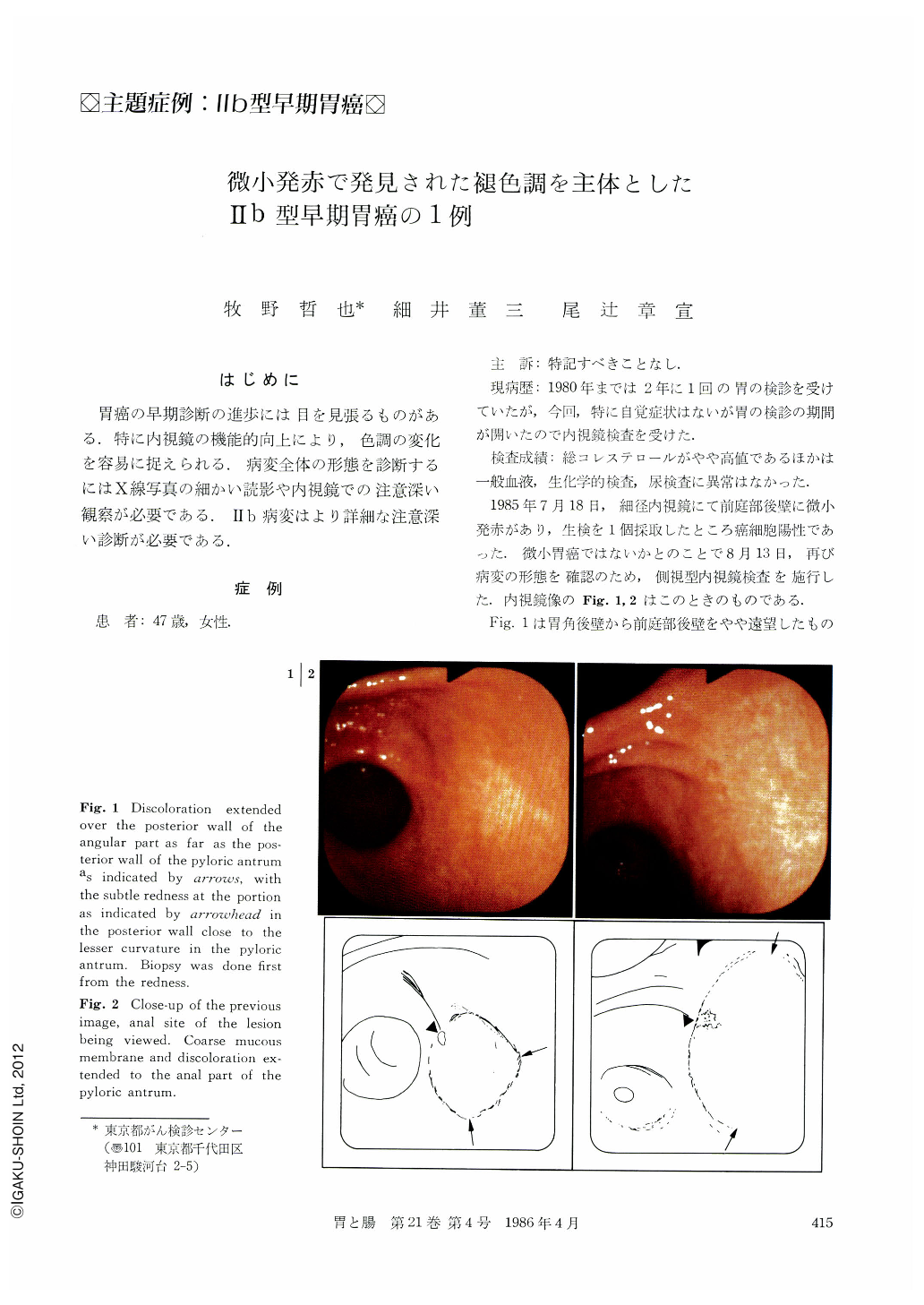
Then, subsequent examination by panendoscopy was performed and cancer cells were again found in the surrounding area. The lesion was flat and not immediately recognized, It was diagnosed as type Ⅱb carcinoma. Endoscopically, discoloration extended over the posterior wall of the angular part as far as the posterior wall of the pyloric antrum, with the subtle redness at the portion in the posterior wall close to the lesser curvature of the pyloric antrum. Mucous membrane looked very coarse in the anal area close to the redness, showing no undulation.
Radiologically the lesion showed a granular appearance which differed from the surrounding area in size and the extent. The border of the lesion could not be determined, though subtle abnormality might be inferred from the area of the lesion.
The entire lesion was situated adjacent to the Fline, measuring 2.5×2.5cm, poorly differentiated cancer being confined in the mucous membrane, con-firming the diagnosis of type Ⅱb.

Copyright © 1986, Igaku-Shoin Ltd. All rights reserved.


