Japanese
English
- 有料閲覧
- Abstract 文献概要
- 1ページ目 Look Inside
はじめに
近年,X線診断学および内視鏡検査は著しく進歩したが,表面平坦型Ⅱb型早期胃癌は現在においてもなお診断困難な病変である.組織学的に5mm未満の大きさを示す微小Ⅱbについては,癌の存在診断そのものが問題となるが,表層拡大型のⅡb病変ではその拡がりが正しく診断されることが,外科治療の面から特に重要である.われわれはX線検査により粘膜の異常を指摘され,内視鏡検査および胃生検にて術前に診断しえた表層拡大型のⅡb型早期胃癌の症例を経験したので報告する.
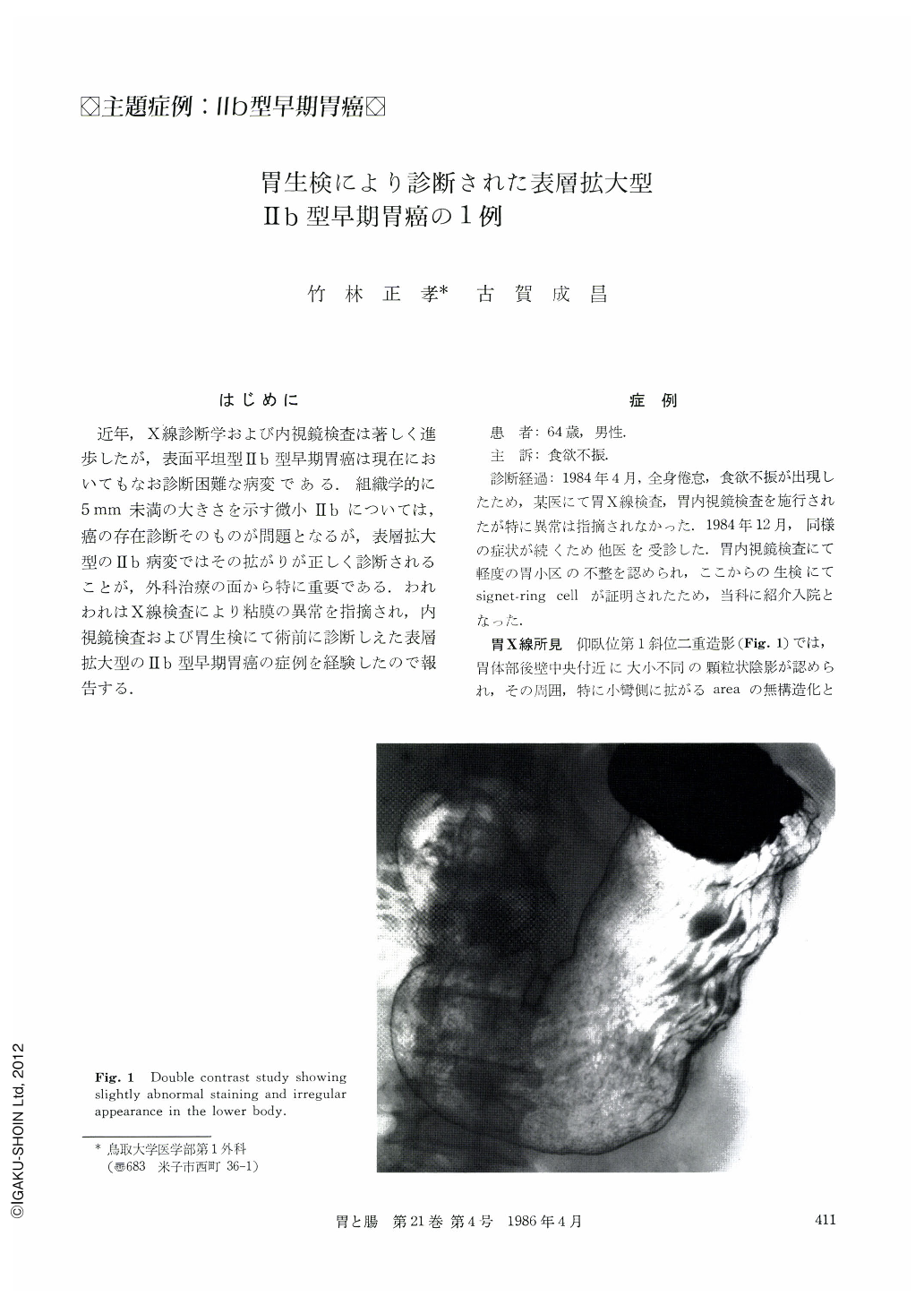
A 64 year-old man was admitted to the hospital for operation of the stomach. Double contrast study of upper GI series had shown abnormal staining and irregularly granular appearance in the gastric lower body. Endoscopic findings had also revealed discoloration without fine mucosal patterns and irregular granulation, using dye-contrast method, at the lower body. By means of these examinations, however, it was difficult to conclude that it was a cancer. It was confirmed by biopsy. Under the diagnosis of IIb, subtotal gastrectomy was performed on January 17, 1985. On the resected specimen, the flat IIb lesion had irregular granularity at the posterior wall of the body. But it was difficult to distinguish the lesion from the surrounding normal mucosa and therefore to determine the extent of it. On microscopic examination the cancer lesion was 7.5×4.0cm in size, and was composed of signet-ring cell carcinoma limited to the mucosal membrane.
In this case, it was useful to carefully perform the step biopsy on the oral side to avoid ow (+) at the operatlon.

Copyright © 1986, Igaku-Shoin Ltd. All rights reserved.


