Japanese
English
- 有料閲覧
- Abstract 文献概要
- 1ページ目 Look Inside
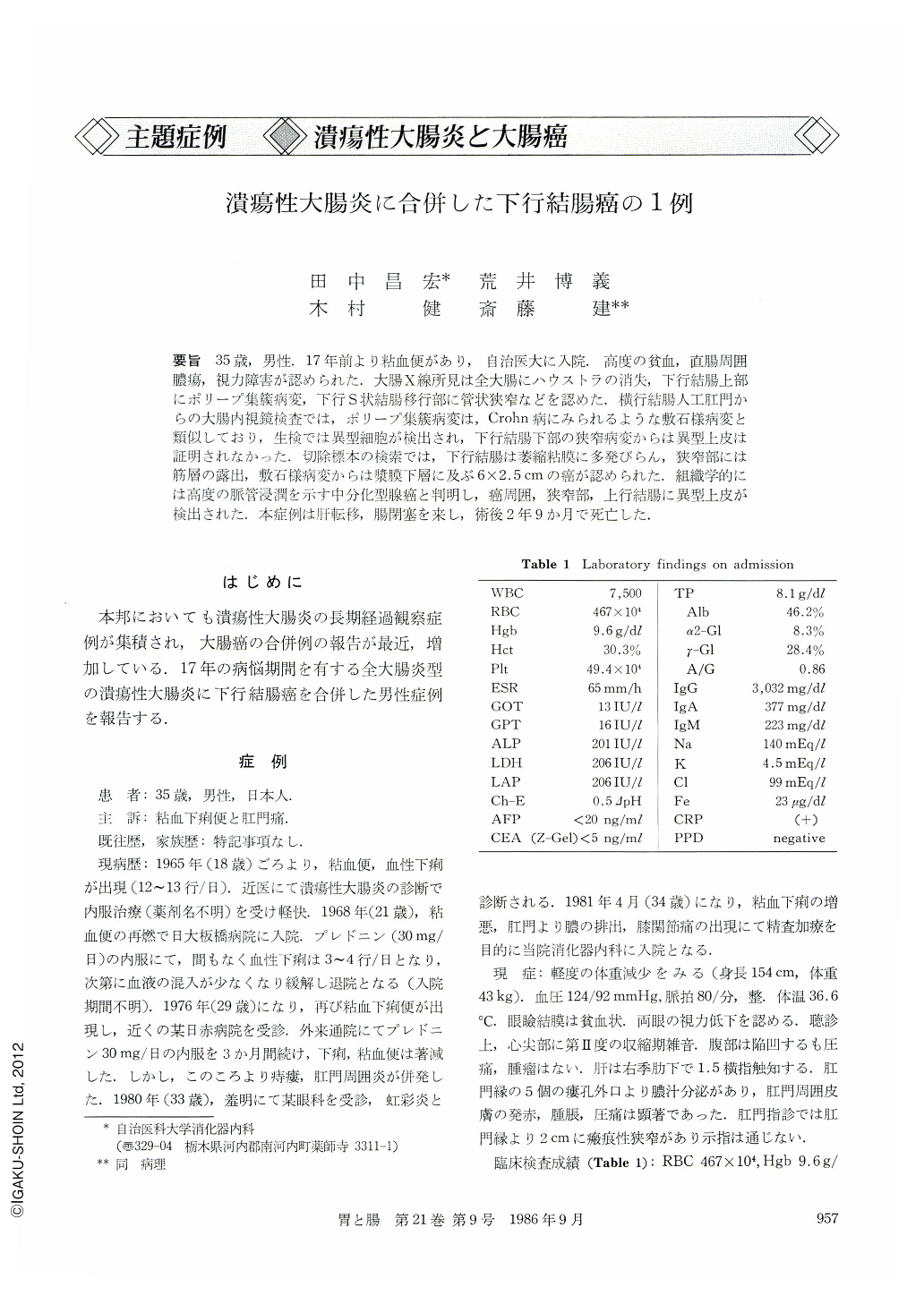
要旨 35歳,男性.17年前より粘血便があり,自治医大に入院.高度の貧血,直腸周囲膿瘍,視力障害が認められた.大腸X線所見は全大腸にハウストラの消失,下行結腸上部にポリープ集籏病変,下行S状結腸移行部に管状狭窄などを認めた.横行結腸人工肛門からの大腸内視鏡検査では,ポリープ集簇病変は,Crohn病にみられるような敷石様病変と類似しており,生検では異型細胞が検出され,下行結腸下部の狭窄病変からは異型上皮は証明されなかった.切除標本の検索では,下行結腸は萎縮粘膜に多発びらん,狭窄部には筋層の露出,敷石様病変からは漿膜下層に及ぶ6×2.5cmの癌が認められた.組織学的には高度の脈管浸潤を示す中分化型腺癌と判明し,癌周囲,狭窄部,上行結腸に異型上皮が検出された。本症例は肝転移,腸閉塞を来し,術後2年9か月で死亡した.
A 35 year-old man with frequent mucobloody stool over a period of seventeen years was admitted to Jichi Medical School Hospital. Severe anemia, periproctal abscess and weakness of sight were noted. Barium enema revealed lack of haustration in the entire colon, polypoid lesion in the descending colon, and tubular narrowing in the descendicosigmoid junction. Endoscopic examination from transverse colostomy showed polypoid lesion of descending colon. The lesion had the cobblestone pattern appearance characteristic of Crohn's disease. Biopsy specimen under direct vision showed marked inflammation with epithelial atypia in the “cobblestony” lesion, and atrophic mucosa without atypia at the descendicosigmoid junction. Resected specimen revealed the atrophic flat mucosa with multiple erosions in the colon on the left side, narrowed segment with exposure of proper muscle coat at the descendicosigmoid junction and advanced cancer (6. 0 X 2. 5 cm) invading the subserosa in the upper part of the descending colon. Histological findings disclosed moderately differentiated adenocarcinoma with marked venous and lymphatic invasion, and various degrees of epithelial dysplasia in the mucosa of the ascending colon and the narrowed portion. Two years and nine months after surgery, the patient died of cancer recurrence, such as liver metastasis and intestinal obstruction.

Copyright © 1986, Igaku-Shoin Ltd. All rights reserved.


