Japanese
English
- 有料閲覧
- Abstract 文献概要
- 1ページ目 Look Inside
要旨 患者は58歳の女性.イレウスの症状を呈し,腹部単純X線像にて左上腹部に水平像を伴った小腸ガス像を認め,上部消化管造影で小腸の完全閉塞像を認めた.手術所見では,Treitz靱帯より肛門側約1.5mの部に順行性の腸重積が認められ,これを整復するに重積部に一致し,くるみ大の腫瘤を認めた.腫瘤を含み腸切除術を施行した.腫瘤の大きさは3×3×2.8cmでポリープ状を示し,表面は赤褐色で軽い出血とびらんが認められ,割面は黄白色を呈した.病理組織学的には,腫瘤は主として粘膜表層から粘膜下層にかけて存在し,一部筋層を巻き込んでいた.周囲組織との境界は比較的明瞭であるが,被膜はみられなかった.腫瘍細胞は比較的大型で卵円形,多角形および長紡錘形で胞体には多数の特徴的な好酸性の微細顆粒が認められた.胞体内顆粒はSudan black B染色,Oil red O染色で強陽性,PAS染色およびS-100蛋白染色でそれぞれ弱陽性,Alcian blue染色で陰性であった.抗S-100蛋白抗体を用いた免疫組織学的検索では明らかな陽性反応が得られなかったが,電顕的には顆粒はautophagosomeあるいはsecondary lysosomeと考えられる顆粒細胞腫で報告されている形態が観察された.以上の所見より顆粒細胞腫と診断し,しかもatypicalな核を有する点,mitosisを認めることより,組織学的には悪性の可能性もあると老えた.患者は術後2年6か月を経た現在,健在である.
Granular cell tumor occurs especially in the soft tissue and the tongue. And the gastrointestinal tract is one of the extremely uncommon locatlons for it We experienced one case of granular cell tumor of the jejunum which intussuscepted. To our knowledge, this is the first reported case in our country.
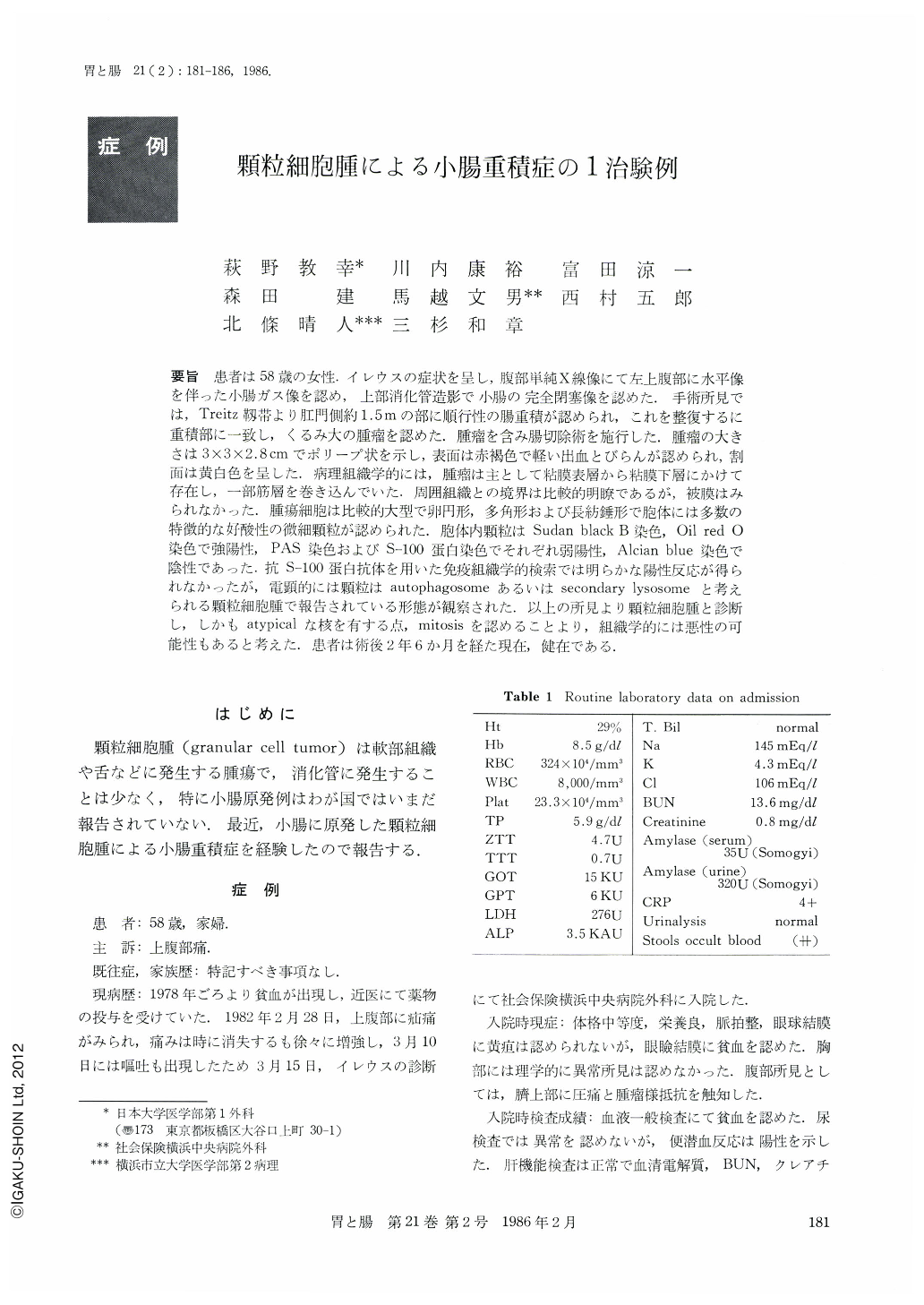
The patient was 58-year-old woman who was admitted to our hospital complaining of intermittent upper abdominal pain with vomiting. The flat plate of the abdomen revealed coils of small intestine disiended by gas and niveau. Upper gastrointestinal series showed complete obstruction of the small intestine. At operation the jejunum intussuscepted. This was reheved and on exploring, a tumor about the size of a walnut was found one hundred and fifty centimeters below the ligament of Treitz. The segment of the small intestine with the tumor was resected. The excised tumor was polypoid, measuring 3×3×2.8cm. The mucosal surface showed erosions with hemorrhage. The cut surface was yellowtsh-white.
On histologic examination the tumor was seen to consist of granular cells. The nests of granular cells were situated between the mucosa and the submucosal collagenous tissue. In the deeper part of the tumor, the granular cell seemed to infiltrate into the muscle layer. The tumor was without capsule. The tumor cells were large and polyhedral, oval or slender spindle-shaped with finely eosinophilic granular cytoplasm. The fine granules of the tumor cells strongly stained by Sudan black B, Oil red O and were faintly colored with PAS and S-100 protein stains. They reacted negatively with Alcian blue. By electron microscopic examination the granules were considered to be autophagosome or secondary lysosome. The tumor had cellular atypism with several mitosis.
We concluded this tumor was granular cell neoplasm with malignant potential.
Copyright © 1986, Igaku-Shoin Ltd. All rights reserved.