Japanese
English
- 有料閲覧
- Abstract 文献概要
- 1ページ目 Look Inside
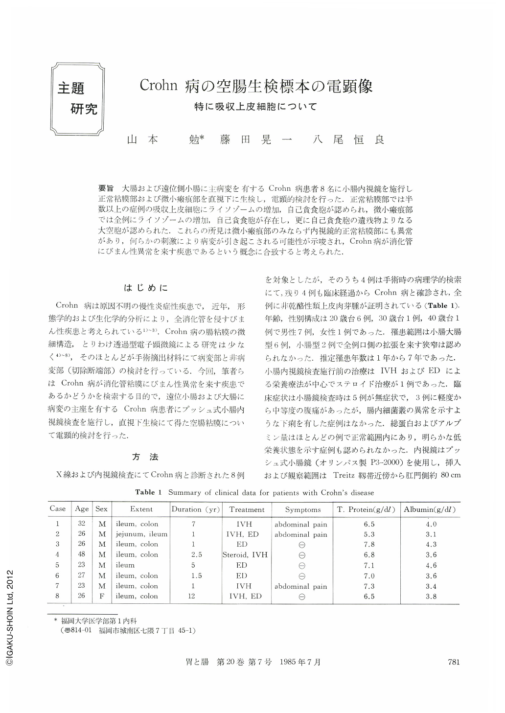
要旨 大腸および遠位側小腸に主病変を有するCrohn病患者8名に小腸内視鏡を施行し正常粘膜部および微小瘢痕部を直視下に生検し,電顕的検討を行った.正常粘膜部では半数以上の症例の吸収上皮細胞にライソゾームの増加,自己貧食胞が認められ,微小瘢痕部では全例にライソゾームの増加,自己貧食胞が存在し,更に自己貧食胞の遺残物よりなる大空胞が認められた.これらの所見は微小瘢痕部のみならず内視鏡的正常粘膜部にも異常があり,何らかの刺激により病変が引き起こされる可能性が示唆され,Crohn病が消化管にびまん性異常を来す疾患であるという概念に合致すると考えられた.
An electron microscopic study was performed to investigate the ultrastructural abnormality in the apparently normal mucosa and minute scar area in eight patients with Crohn's disease. Biopsy specimens were taken under direct vision during small intestinal endoscopy (normal mucosa: eight cases, scar area: two cases). The discernible alterations were described as follows.
1) Apparently normal mucosa: Supranuclear portion of the epithelial cells showed an increase in heterogenous lysosomes in five cases. Autophagic vacuoles and residual bodies were frequently seen in the perinuclear and the lateral portions of the epithelial cells in four cases. Myelin-like structures, presumably residual bodies, were also seen in the cytoplasm and the intercellular spaces.
2) Minute scar area: In addition to the findings concerning the increased number of lysosomes and frequent occurrence of autophagic vacuoles similar to those found in the normal mucosa, various sized vacuoles containing degenerated cell organella were observed in the apical portion. Therefore, some of them were considered to be residual bodies not yet discharged into the extracellular space. These vacuoles were relatively rare in the epithelial cell of the normal mucosa. Neither viral particles nor degraded bacteria were detected in the normal mucosa and the minute scar area.
The autophagic vacuoles in the small intestinal epithelium have been reported in various pathological conditions such as starvation, x-ray irradiation and marasmus. The present cases have no such clear cause for cell injury. Therefore, our findings of autophagic vacuoles and residual bodies even in the normal mucosa may possibly be explained as a consequence of the intestinal lesion of Crohn's disease. And the epithelial cell necrosis, so called“patchy necrosis”, would be caused when these structures are ruptured by noxious agents from the lumen.
These findings, above mentioned, are compatible with the concept of Crohn's disease as a diffuse lesion of the gastrointestinal tract.
Copyright © 1985, Igaku-Shoin Ltd. All rights reserved.