Japanese
English
- 有料閲覧
- Abstract 文献概要
- 1ページ目 Look Inside
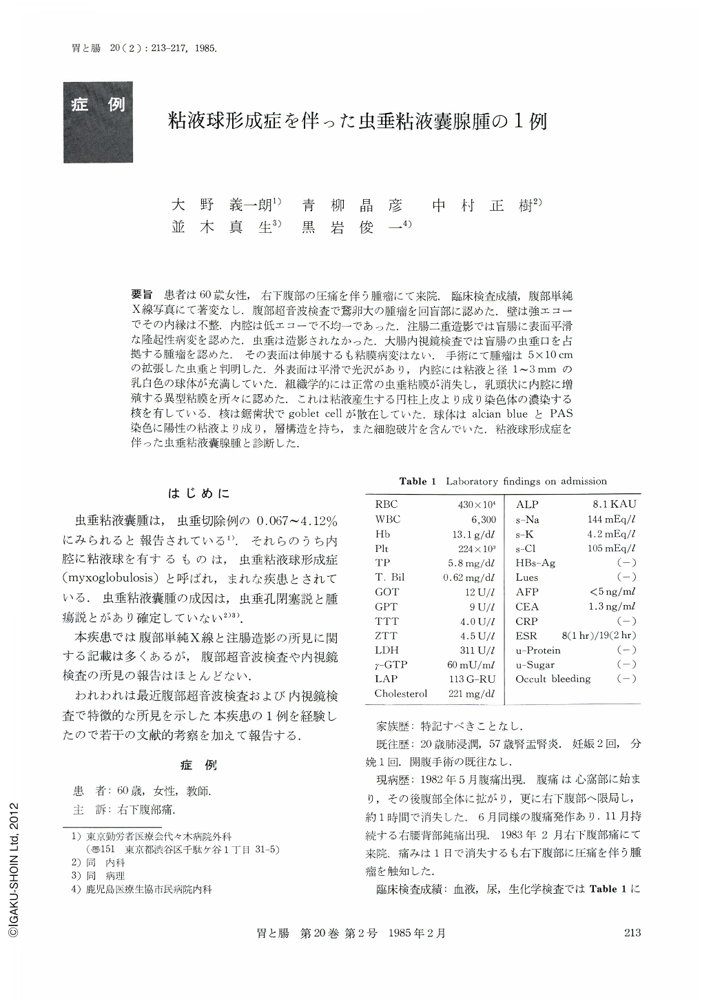
要旨 患者は60歳女性,右下腹部の圧痛を伴う腫瘤にて来院.臨床検査成績,腹部単純X線写真にて著変なし,腹部超音波検査で鷲卵大の腫瘤を回盲部に認めた.壁は強エコーでその内縁は不整.内腔は低エコーで不均一であった.注腸二重造影では盲腸に表面平滑な隆起性病変を認めた.虫垂は造影されなかった.大腸内視鏡検査では盲腸の虫垂口を占拠する腫瘤を認めた.その表面は伸展するも粘膜病変はない.手術にて腫瘤は5×10cmの拡張した虫垂と判明した.外表面は平滑で光沢があり,内腔には粘液と径1~3mmの乳白色の球体が充満していた.組織学的には正常の虫垂粘膜が消失し,乳頭状に内腔に増殖する異型粘膜を所々に認めた.これは粘液産生する円柱上皮より成り染色体の濃染する核を有している.核は鋸歯状でgoblet cellが散在していた.球体はalcian blueとPAS染色に陽性の粘液より成り,層構造を持ち,また細胞破片を含んでいた.粘液球形成症を伴った虫垂粘液嚢腺腫と診断した.
A 60 year-old woman was admitted because of a right lower quadrant mass and tenderness. Laboratory studies revealed no abnormalities such as occult bleeding, inflammation signs. A plain film of abdomen showed no abnormality. An ultrasound scan demonstrated a goose-egg sized mass in the ileocecal portion. The wall was echogenic and its inner line was irregular. The lumen was hypoechoic, but not homogeneous. Double contrast study of the colon revealed a smooth extrinsic mass in the caecum. The appendix was not filled. Colonoscopy visualized a tumor in the caecum occupying the orifice of the appendix. The surface was expanded without mucosal destruction.
At surgery, the mass was an enlarged appendix, 5 cm in diameter and 10 cm in length. The external surface was smooth and glistening. The lumen was filled with mucus and milky-white globular bodies from 1 to 3 mm in diameter.
Histologically, the normal appendiceal mucosa was absent. Papillary projections were partly seen in the lumen. These processes were lined by tall columnar mucus-secreting cells with basal hyperchromatic nuclei: Pseudostratification was noted and goblet cells were scattered. The globules were consisted of alcian-blue and PAS positive mucin, forming concentric layer and cell debris.

Copyright © 1985, Igaku-Shoin Ltd. All rights reserved.


