Japanese
English
- 有料閲覧
- Abstract 文献概要
- 1ページ目 Look Inside
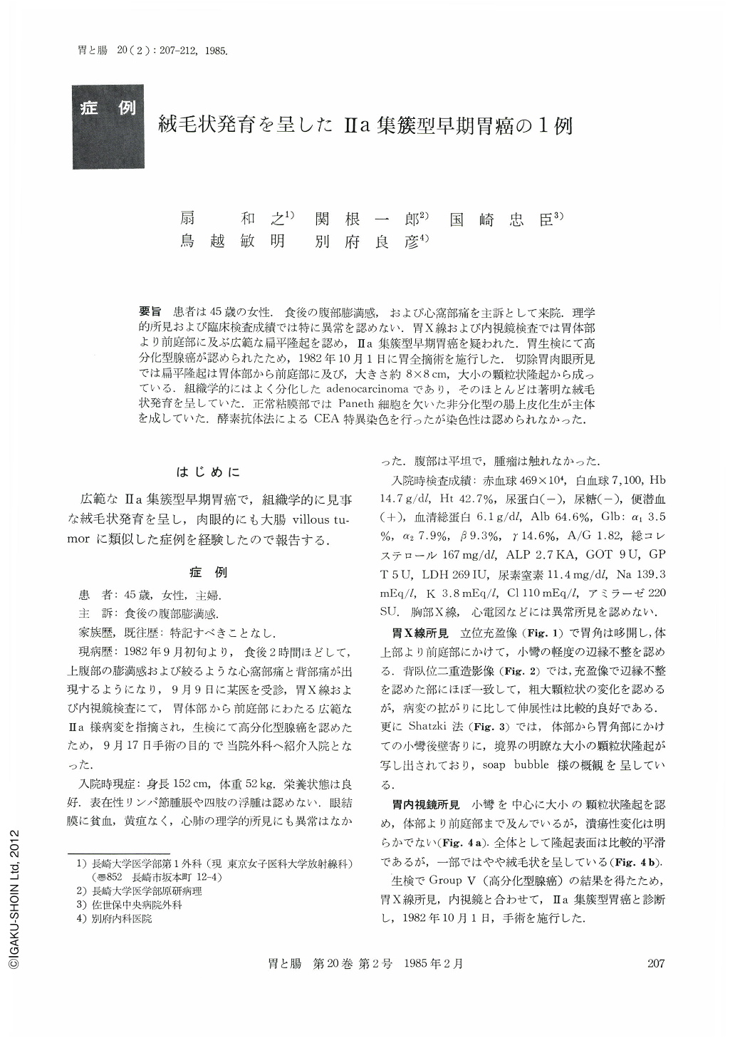
要旨 患者は45歳の女性.食後の腹部膨満感,および心窩部痛を主訴として来院.理学的所見および臨床検査成績では特に異常を認めない.胃X線および内視鏡検査では胃体部より前庭部に及ぶ広範な扁平隆起を認め,Ⅱa集簇型早期胃癌を疑われた.胃生検にて高分化型腺癌が認められたため,1982年10月1日に胃全摘術を施行した.切除胃肉眼所見では扁平隆起は胃体部から前庭部に及び,大きさ約8×8cm,大小の顆粒状隆起から成っている.組織学的にはよく分化したadenocarcinomaであり,そのほとんどは著明な絨毛状発育を呈していた.正常粘膜部ではPaneth細胞を欠いた非分化型の腸上皮化生が主体を成していた.酵素抗体法によるCEA特異染色を行ったが染色性は認められなかった.
A 45 year-old woman with abdominal fullness and epigastric pain after meal visited our clinic. Physical examinations and laboratory data showed no particular abnormalities. Barium meal and endoscopical examinations revealed a flat elevated lesion of the gastric body down to the pyloric antrum, which was suspected as Ⅱa type early cancer. Since gastric biopsy indicated well differentiated adenocarcinoma, total gastrectomy was performed on October 1, 1982. The resected stomach showed a flat elevated lesion with granular and villous surface, measuring about 8×8 cm, located in the gastric body and pyloric antrum.
Histology showed well differentiated adenocarcinoma with remarkable villous pattern. The surrounding mucosa was occupied by intestinal metaplasia which lacks of Paneth cells.
CEA staining using immuno-peroxidase method was negative.

Copyright © 1985, Igaku-Shoin Ltd. All rights reserved.


