Japanese
English
- 有料閲覧
- Abstract 文献概要
- 1ページ目 Look Inside
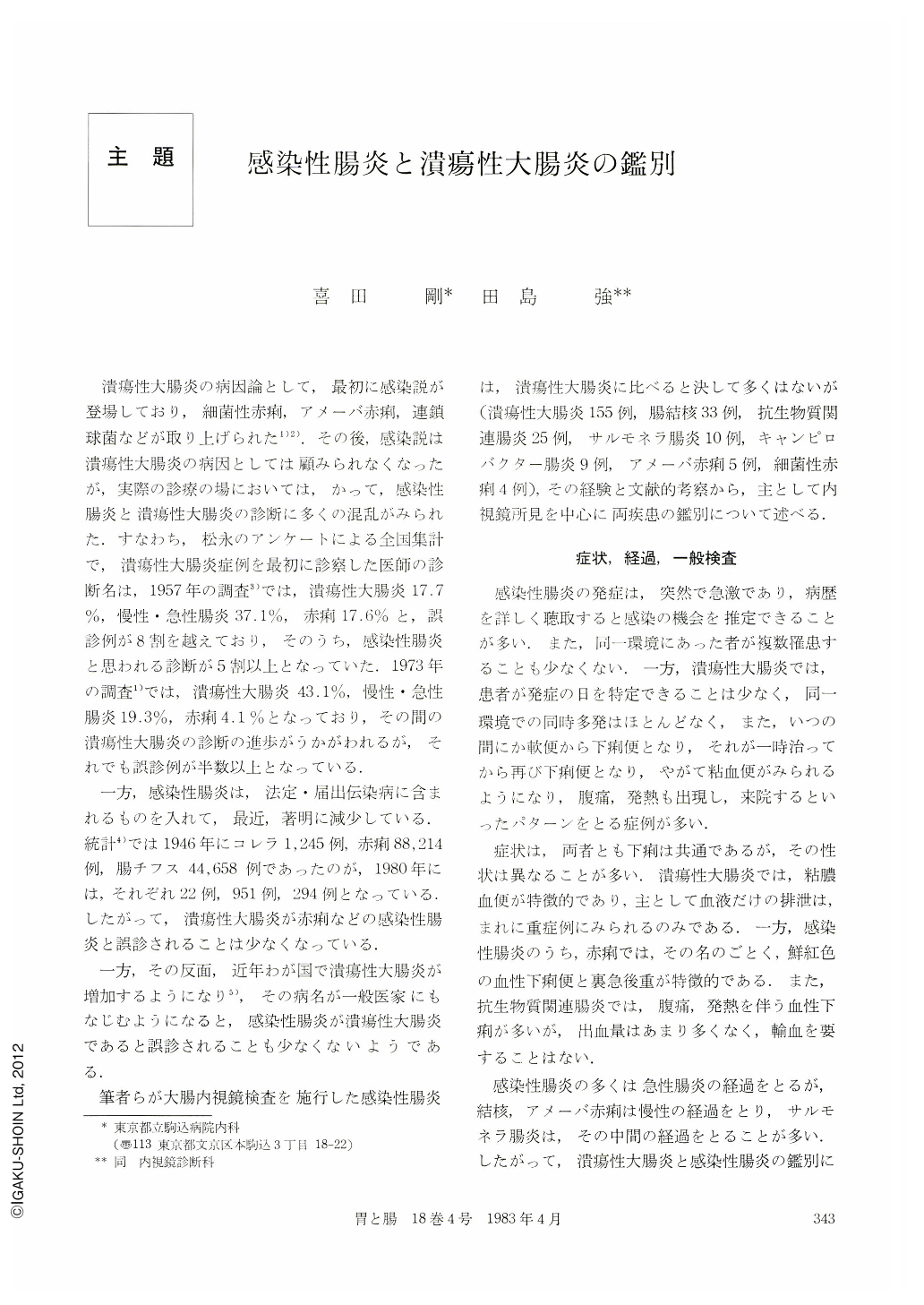
潰瘍性大腸炎の病因論として,最初に感染説が登場しており,細菌性赤痢,アメーバ赤痢,連鎖球菌などが取り上げられた.その後,感染説は潰瘍性大腸炎の病因としては顧みられなくなったが,実際の診療の場においては,かって,感染性腸炎と潰瘍性大腸炎の診断に多くの混乱がみられた.すなわち,松永のアンケートによる全国集計で,潰瘍性大腸炎症例を最初に診察した医師の診断名は,1957年の調査では,潰瘍性大腸炎17.7%,慢性・急性腸炎37.1%,赤痢17.6%と,誤診例が8割を越えており,そのうち,感染性腸炎と思われる診断が5割以上となっていた.1973年の調査では,潰瘍性大腸炎43.1%,慢性・急性腸炎19.3%,赤痢4.1%となっており,その間の潰瘍性大腸炎の診断の進歩がうかがわれるが,それでも誤診例が半数以上となっている.
一方,感染性腸炎は,法定・届出伝染病に含まれるものを入れて,最近,著明に減少している.統計では1946年にコレラ1,245例,赤痢88,214例,腸チフス44,658例であったのが,1980年には,それぞれ22例,951例,294例となっている.したがって,潰瘍性大腸炎が赤痢などの感染性腸炎と誤診されることは少なくなっている.
Most of the infections of the intestines except amebic colitis and tuberculous enterocolitis have abrupt onset and take acute self-limited course. On the other hand, ulcerative colitis has gradual or insidious onset and pursues chronic course.
Basic endoscopic feature of ulcerative colitis is diffuse inflammation that usually involves the rectum.
Stool culture for Campylobacter fetus ss. jejuni has become possible from the beginning of 1980 at our hospital. Since then, we have experienced nine cases with Campylobacter infection of the intestines. Colonic abnormalities were present in seven among these nine cases. Aphthoid ulcers were seen in two cases. Small patchy reddenings were seen in four cases. Consequently, in a total of six cases, tiny abnormalities with spotty distribution were seen. But, in one case, diffuse changes were seen 〔Case 3, Fig. 3, 4〕 and were indistinguishable from ulcerative colitis endoscopically.
Endoscopic findings of Shigella dysentery were patchy to tiny reddenings, small erosions and aphthoid ulcers. Mucosa between these inflammations was intact and these findings were apparently different from those of ulcerative colitis.
We found out colonic lesions in eight cases among a total of 10 cases with Salmonella infection of the intestines. Diffuse reddening was seen segmentally in three cases. And patchy reddenings were seen in another three cases. Aphthoid ulcers only were seen in one case. And in one case, serial changes of diffuse reddening to broad longitudinal ulcer, and finally to linear ulcer scar were seen.
Stool culture of Clostridium difficile became posible since August 1982 at our hospital. Before that period, we have experienced 18 cases with antibiotic-associated colitis. Culture of stool and/or biopsy specimen was positive for Klebsiella oxytoca in 10 cases and was negative in eight cases. Endoscopic characteristics of Klebsiella oxytoca positive cases were linear, patchy and diffuse reddening, erosions and bleeding. No obvious endoscopic characteristics except two cases of pseudomembrane formation were seen among the cases with antibiotic-associated colitis with negative stool culture. During the period when the stool culture for Clostridium difficile have become possible, we have experienced seven cases with antibiotic-associated colities. Clostridium dificille was isolated in five among these seven cases. Endoscopic findings of Clostridium difficile positive cases were small reddenings and ulcers in four cases and one case showed diffuse reddening and erosions from the rectum to the descending colon and the endoscopic findings of this case resembled those of ulcerative colitis.
Endoscopic abnormalities of tuberculous colitis were distributed most frequently at the ileocecal area. Skip lesions with circular ulcer, convergency, concentric stenosis and small inflammatory polyps were characteristic endoscopic findings.
Four among five cases with amebic colitis showed varioliform erosions and ulcers. And these findings improved markedly after therapy with metronidazole.
Pattern of distribution of endoscopically observed lesions and each item of endoscopic findings of infections of the intestines were different from those of ulcerative colitis in most cases and each infection of the intestines has its characteristics in endoscopic features. But some cases with infections of the intestines could not be differentiated from ulcerative colitis by endoscopic features alone, thus close scrutiny and follow-up were considered to be necessary in dealing with patients with colonic inflammation.

Copyright © 1983, Igaku-Shoin Ltd. All rights reserved.


